
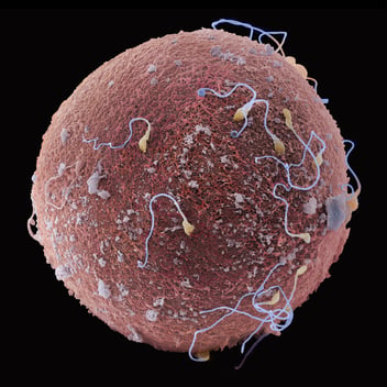
Electron microscopy enables researchers to study biological structures at much higher resolution than conventional light microscopy, from cell and tissue surfaces to organelles, membranes and molecular complexes.
Listen to article
In life sciences, obtaining a high-resolution image is often only the beginning. The real challenge is understanding the biological story it tells.
How does a virus enter a cell and reorganize its internal architecture?
How do neurons connect into functional networks?
How do therapeutic interventions alter cellular organization?
And how do molecular machines interact within their biological environment?
For decades, microscopy has helped scientists reveal what was once invisible. Every improvement in resolution has uncovered new levels of biological complexity, from tissues and cells to organelles and molecular assemblies.
Yet seeing more detail does not automatically mean understanding more biology.
A high-resolution image may show what a structure looks like, but understanding its role often requires a broader view.
Where is it located?
What surrounds it?
How does it interact with neighbouring structures?
And how does it contribute to the function of the cell, tissue or organism as a whole?
This is where electron microscopy becomes more than a tool for imaging. It helps researchers connect structure with function, and individual observations with biological context.
From Seeing Structures to Understanding Their Context
Different electron microscopy approaches offer different windows into biological organization.
Life sciences microscopy is as diverse as biology itself. A butterfly wing, a plant root, a bacterial biofilm, a cultured cell and a resin-embedded brain section may all end up in an electron microscope, yet each presents a different challenge and may require a different workflow.
The first challenge often appears before imaging even begins: preserving the biological information researchers want to study.
Some specimens benefit from conductive coating, which helps reduce charging effects and reveal fine surface details. Others are too delicate to modify and require imaging conditions that minimize sample preparation. When researchers need to investigate what lies beneath the surface, cells and tissues can be preserved in resin, creating stable samples for ultrastructural analysis.
Choosing the right workflow is therefore just as important as choosing the right microscope.
This is where Tescan Life sciences solutions are designed to support researchers across the journey from sample to insight. By combining SEM, FIB-SEM and cryogenic workflows, Tescan helps laboratories adapt their imaging approach to the biological question, the sample type and the level of detail required.
The goal is not simply to produce a beautiful image, but to preserve the structures that matter and extract meaningful biological information from them.
From Surface Morphology to Cellular Architecture
Scanning electron microscopy (SEM) reveals the morphology and surface architecture of cells, tissues, whole organisms and other biological specimens. SEM provides a versatile way to investigate biological surfaces, from cultured cells and microorganisms to plants, insects, tissues and biomaterials.
Depending on the sample and research question, researchers may image conductive-coated specimens, uncoated samples under low-vacuum conditions, or cryo-preserved and freeze-fractured specimens that retain their surface morphology.
SEM can reveal how cells spread across a surface, how tissues develop complex architectures, how microorganisms organize into communities, how plants adapt their structures to their environment, or how biological materials interact with surroundings.
For surface-focused questions, this flexibility allows researchers to choose preparation and imaging conditions that match the specimen while preserving the morphological information they need.


But many biological structures cannot be fully understood from a single image.
Organelles form interconnected networks, membranes create complex interfaces, and tissues extend across dimensions that are impossible to capture in a two-dimensional view. Understanding how these structures are organized requires the ability to follow them through a volume.
From 2D Images to 3D Biology
Three-dimensional electron microscopy offers several approaches to this challenge, each balancing imaging volume, resolution and workflow flexibility in a different way.
Array Tomography (AT) uses a standalone ultramicrotome to generate serial ultrathin sections that are collected on a substrate and then imaged individually in the SEM to create a series of 2D images, which are then reconstructed in the Tescan 3D Viewer™ to create a 3D reconstruction. Because the physical sections remain available after imaging, researchers can revisit the sample, perform additional analyses and correlate information across different imaging modalities.

In Serial Block-Face SEM (SBF-SEM), sectioning is integrated directly into the microscope. An in-chamber ultramicrotome automatically removes thin layers from the resin block between imaging cycles, exposing a new surface for acquisition. After each layer is removed, the newly exposed surface is imaged, generating a stack of 2D images that is then reconstructed into a 3D volume. This highly automated workflow enables efficient reconstruction of large cellular and tissue volumes, making it well suited to applications such as connectomics, developmental biology and tissue architecture studies. Because material is removed during acquisition, however, the process is inherently destructive.

When the focus shifts from larger volumes to finer ultrastructural detail, FIB-SEM tomography offers a different perspective.
FIB-SEM tomography benefits from the focused ion beam’s ability to precisely mill very thin slices of material from the sample, exposing a fresh surface for SEM imaging after each milling step. This precise ion-beam control enables the generation of three-dimensional datasets with near-isotropic resolution, providing comparable levels of detail across the X, Y and Z dimensions.
While the accessible volume is typically smaller than that of SBF-SEM or Array Tomography, the higher Z-resolution can be transformative. Researchers can trace mitochondrial networks, membrane contacts, synaptic architectures and intracellular interfaces through a volume, revealing nanoscale relationships that may be difficult to resolve in conventional serial sections.
FIB-SEM therefore provides a bridge between three-dimensional imaging and ultrastructural analysis, offering a detailed view of how cellular structures are organized in space.

Moving Closer to Native Biology
All these workflows can provide remarkable insights into cellular and tissue organization. However, they typically rely on coating, chemical fixation, dehydration, staining or resin embedding. These preparation steps make biological specimens stable and compatible with electron microscopy, but they can also alter aspects of their original organization.
As researchers explore biology at one scale, their observations often lead to questions at another. Understanding how cells are organized opens the door to questions about individual organelles, while studying organelles reveals the need to understand the molecular complexes and machines that drive their function.
To explore biology at this level, researchers need approaches that preserve biological structures and their surrounding context as close to their original state as possible.
This is where cryo-electron microscopy (cryo-EM) has transformed structural biology.
Cryo-EM addresses this challenge through vitrification - a rapid freezing process that prevents the formation of ice crystals and preserves biological structures in a state as close to their native organization as possible.
One of the major breakthroughs came with single-particle analysis (SPA), which enabled researchers to determine the structures of proteins, viruses and molecular complexes at near-atomic resolution in the transmission electron microscope (TEM). For the first time, molecular machines could be studied in extraordinary structural detail.
But, as often happens in biology, every answer created new questions.
Knowing what a molecular complex looks like is important. Understanding where it is located, what surrounds it and how it interacts with other cellular structures can reveal another layer of biological meaning.
This has driven the rapid adoption of cryo-electron tomography (cryo-ET) which moves beyond isolated molecular views by enabling molecular complexes and cellular structures to be visualized within thin regions of cells and in their surrounding biological environment.
Because intact cells and tissues are often too thick for electron-transparent imaging, researchers use cryo-FIB milling to prepare thin lamellae from vitrified samples. This provides access to selected regions deep within the specimen while retaining their cellular context for subsequent cryo-ET investigation.
The role of FIB-SEM does not end with lamella preparation.
The same platform can also support cryo-volume imaging, extending the field of view from individual molecular complexes and targeted regions to larger volumes of vitrified cells and tissues, similar to FIB-SEM tomograph in the room temperature described earlier.
This adds another level of biological context. Researchers can investigate how molecular machinery relates to organelles, how cellular structures are organized throughout a volume, and how local molecular detail fits into the larger architecture of the cell.


Connecting the Scales of Biology
Sometimes researchers need to see the whole specimen before deciding where to look closer. Sometimes delicate surface structures require carefully optimized imaging conditions. Sometimes the critical information lies beneath the surface, where three-dimensional imaging can reveal hidden organization. And sometimes preserving the sample as close to its original state as possible is essential to understanding what happens at the cellular or molecular level.
The most useful workflow is therefore the one that matches the sample, the scale and the biological question.
At Tescan, our Life sciences solutions are shaped through close interaction with researchers, core facilities, application specialists and technology developers. From surface morphology and low-kV imaging to volume EM, FIB-SEM tomography and cryo-FIB-SEM workflows for cryo-ET and cryo-volume imaging, our aim is to provide researchers with the flexibility to move between scales and perspectives.
Biological insight rarely comes from a single image. It comes from connecting what we see at different resolutions - from overall organization to ultrastructural detail, from surfaces to volumes, and from molecular machinery to the cellular environment in which it exists.
Understanding biology means seeing these connections.
And sometimes, the way to understand the smallest details is to start with the bigger picture.
Written by Jana Šmídová
Product Marketing Manager, Tescan


Related Resources
Life Sciences Solutions
Explore microscopy workflows from cellular imaging to cryo-EM.

Cryo-EM & Correlative Workflows
Discover workflows for challenging biological samples.

Cryo-ET Sample Preparation
Explore cryo-EM workflows for bacteria–biomaterial interfaces.