Tescan Picannto   

AI-assisted 3D annotation and segmentation software 

Picannto - hero - desktop kopie
Picannto - hero - mobile kopie

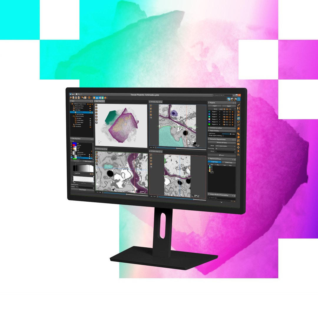
Tescan Picannto™ extends the Tescan 3D Viewer™ workflow with dedicated 3D annotation and segmentation capabilities. It helps users turn complex volumetric datasets into structured, quantitative outputs while keeping visualization, segmentation, and presentation connected within the Tescan 3D software ecosystem.

Designed specifically for 3D segmentation workflows, Tescan Picannto™ combines manual precision tools, semi-automatic methods, and AI-assisted segmentation in one purpose-built environment. Users can define, refine, and validate regions of interest with greater speed and control, without switching between disconnected software tools.

Advanced AI-assisted tools, including methods based on Segment Anything Model 2, help accelerate object detection and region definition in complex datasets. Users remain in control of the final result and can combine AI output with manual refinement and validation to create precise and reproducible segmentations.

Built on a high-performance 3D rendering engine, Tescan Picannto™ enables smooth interaction with large volumetric datasets and supports a wide range of 3D and medical imaging data formats. Its task-oriented interface helps users focus on segmentation logic and analysis rather than software operation.

Together with Tescan 3D Viewer™, Tescan Picannto™ creates a connected workflow from preprocessing and visualization to segmentation, overlays, and presentation. Enhanced datasets can be transferred from Tescan 3D Viewer™ to Tescan Picannto™ for segmentation, while segmented regions can be exported as NumPy files for downstream analysis, machine learning, validation, and reproducible workflows.

Tescan Picannto™ is also extensible through Compox™, the Python-based extensibility layer. Users can connect custom algorithms, AI models, and specialized segmentation workflows directly to the application, while developing methods in their own Python environment and controlling them from the familiar Tescan Picannto™ interface.

This makes Tescan Picannto™ more than a conventional segmentation tool: it is a connected, AI-assisted, and extensible segmentation environment that can adapt to specific materials, structures, research methods, and industrial analysis requirements. 

Typical applications

  • Advanced 3D segmentation
    Segment complex volumetric datasets using manual, semi-automatic, and AI-assisted tools. 


  • Life science and medical image annotation
    Segment and annotate diverse 3D imaging datasets for research, validation, quantitative analysis, and machine learning workflows.

  • AI model development and validation 
    Create annotated datasets and export segmented regions as NumPy files for machine learning, benchmarking, and reproducible research. 

  • Custom segmentation workflows with Compox™  
    Extend Tescan Picannto™ with proprietary algorithms, AI models, or specialized segmentation workflows for specific materials, tissues, structures, or analysis requirements. 

WHERE Tescan Picannto™

makes the difference

Purpose-built interface for 3D annotation and segmentation

Tescan Picannto™ provides a task-oriented UI/UX designed specifically for volumetric annotation and segmentation. Users can combine manual refinement, semi-automatic tools, and AI-assisted methods in one focused environment, helping them create precise and reproducible segmentation results with less software complexity. 

AI-assisted segmentation with Segment Anything Model 2

Integrated AI-assisted tools, including methods based on Segment Anything Model 2, help accelerate object detection and region definition in complex 3D datasets. Users remain in control of the final result and can combine AI output with manual refinement and validation when accuracy and reproducibility are critical. 

Smooth interaction with large volumetric datasets

Tescan Picannto™ supports a wide range of volumetric and medical imaging data formats and is built on a high-performance 3D rendering engine for smooth interaction with large datasets. This allows users to work directly with complex 3D data in an environment optimized for annotation and segmentation 

Open and extensible with Compox™

Tescan Picannto™ is not limited to built-in tools. Through Compox™, the Python-based extensibility layer, users can connect custom algorithms, AI models, and specialized processing workflows directly to the Tescan Picannto™.
 
Algorithms can be developed in the user’s own Python environment and exposed through the Tescan Picannto™ interface, so advanced methods can be controlled and executed as part of the standard 3D data workflow. This gives research and industrial teams a practical way to adapt the software to specific materials, methods, and analysis requirements without rebuilding their toolchain.

Tescan Spectral CT Applications

_ Application area icon (3)
Tescan Spectral CT in Electronics and Semiconductors

Non-destructive spectral imaging for material verification and internal inspection of advanced electronic assemblies.

TrueContrast™ multi-energy imaging for differentiating polymers, metals, and encapsulants

  • K-edge detection for accurate identification of high-Z elements

  • Non-destructive visualization of interfaces and buried defects in electronic packages

  • Spectrum comparison tools for verifying material uniformity and contamination sources

Tescan Spectral CT delivers compositional insight where conventional micro-CT cannot. Engineers can distinguish packaging materials, solder alloys, and internal structures without sectioning or coating—supporting reliable failure analysis, design validation, and quality assurance across next-generation electronic devices.

_ Application area icon
Tescan Spectral CT in Materials Science

Multi-energy micro-CT for elemental and structural differentiation in complex, multi-phase materials.

  • Elemental mapping for identifying fillers, dopants, or additives in polymer composites

  • Spectral contrast for distinguishing polymers, ceramics, and metals with similar densities

  • Non-destructive 3D analysis of internal interfaces and phase boundaries

  • Integrated Spectral Suite for correlating elemental and structural data in one workflow

In materials research, Spectral CT enables scientists to explore the relationship between structure and composition without destroying samples. Researchers can visualize distribution of additives, analyze composite uniformity, and study degradation pathways—supporting more reliable material design and performance assessment.

Application area=GS
Tescan Spectral CT in Geoscience and Mining

Full-spectrum micro-CT for 3D elemental and structural insight into geological samples.

  • Non-destructive 3D elemental mapping of rocks, ores, and mineral inclusions

  • K-edge detection for locating and identifying high-value or trace elements like gold or rare earth elements

  • Enhanced contrast for complex mineral assemblages and pore networks

  • Large-volume spectral scanning for full-core or fragment-scale analysis

For geoscientists and mining professionals, Spectral CT provides simultaneous elemental and structural data from intact samples. It supports mineral identification, ore classification, and recovery assessment—reducing dependency on destructive assays while preserving geological material for further study.

TESCAN SPECTRAL SUITE

INTEGRATION THAT DRIVES INSIGHT

Tescan Spectral CT works seamlessly with SPECTRAL Suite and Acquila™ micro-CT control software to streamline spectral imaging from acquisition to analysis. Used as an add-on with the UniTOM XL platform, users can target volumes of interest, capture full-spectrum data, and visualize structural and elemental information in one workflow.

Researchers benefit from intuitive controls, guided reconstruction, and automated spectral processing. Spectral Suite handles spectrum matching, K-edge detection, and compositional mapping with minimal setup. This delivers consistent, reproducible results across samples and accelerates multi-energy micro-CT analysis.

Unlocked content

Unlock
the trial version

leave a contact to access

Get the most out of Tescan

This is more than information; it's an advantage. We've compiled our technical whitepapers, detailed product flyers, and on-demand webinars to provide you with the knowledge that makes a real difference. Sign up now to access the insights you need to make an impact.

Trial Version

The trial version is available for 30 days and includes the full functionality of the application without any feature limitations.

Online registration is required to activate the trial. The computer must remain connected to the internet while the application is in use.

Each user account can activate one trial version. The same limitation applies to each individual computer.

Follow the trial activation instructions:

Download the trial version of Tescan Picannto

GET IN Touch

Contact us

map

Where you can find us:

Tescan Brno
Libušina třída 21
623 00 Brno
Czech Republic


info@Tescan.com 

130405923 us US 37.09024 -95.712891 25.3575 29.349345 20.67957527 42.082797 39.91384763 -33.693421 13.93320106 3.039986586 31.997988 38.050985 47.579533 48.1485965 58.375799 54.663142 19.195447 56.975106 47.916997 50.493053 45.868592 10.79556993 44.35660598 43.2371604 55.536415 14.557577179752773 32.100937 -6.116829 -6.212299277967318 33.600194 -12.08688 23.7104 -33.471062 31.998740087 -23.69149395 43.462349 51.529848 49.1893523 49.197486 25.072375 31.075811 1.299027 40.676979 52.30150662 51.013813 35.684121 37.566531 52.246622 40.581349 39.911632 -26.1811371 41.818215 33.429928

No distributors found.