Cryo-electron microscopy (cryo-EM) has revolutionized structural biology. By enabling the observation of cellular structures in their near-native state at molecular resolution, this groundbreaking technology is reshaping how scientists understand life at the nanoscale. Whether preparing thin lamellae for Cryo-Electron Tomography (Cryo-ET) to obtain the finest structural details or performing Cryogenic volume imaging, Tescan workflows ensure precision, reliability, and sample integrity throughout the entire process.
Preserve Native Biological Structure for Reliable Cryo-ET and Volume Electron Microscopy
Achieving reproducible cryo-ET data starts with precise sample preparation and preserved structural integrity. Tescan solutions deliver cryogenic FIB-SEM workflows that maintain native cell and tissue morphology. This enables confident cryo-lamella preparation, high-resolution surface imaging, and 3D volume reconstruction in life-science research.

FOR THE ENTIRE LIFE SCIENCES RESEARCH AND DEVELOPMENT PROCESS
Cryo-ET Sample Preparation
.webp?width=1264&height=1020&name=2_A%20correlation%20of%20%20SEM%20(1).webp)
Surface Morphology Analysis
Enable reproducible results and reveal delicate biological structures for reliable surface morphology studies. Tescan workflows provide detailed, high-contrast imaging of coated, uncoated, and hydrated samples, revealing essential topographical features that drive discovery in the life sciences.

Volume Electron Microscopy
Understanding biological systems requires more than a single plane of imaging—how organelles interact within cells, how cells organize in tissues, or how microbial communities form in ecological niches. Tescan volume electron microscopy (vEM) provides multi-scale, 3D insights into cells, tissues, and small organisms.

"Tescan AMBER X 2, our latest innovation in materials analysis technology. The AMBER X 2, with its advanced Plasma FIB-SEM capabilities, offers unmatched speed, precision, and versatility, setting a new standard in the field."
PETR KLIMEK
Product Marketing Director Material & Geo Science
Tescan

Used in This Workflow
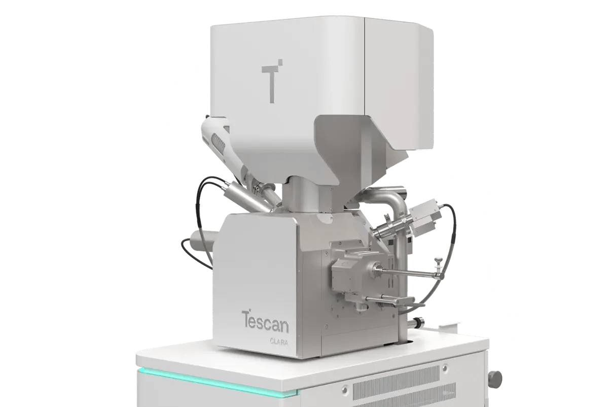
Tescan CLARA™
Tescan CLARA™ is engineered for advanced biological imaging under both ambient and cryogenic conditions. It supports a wide range of workflows, enabling researchers to capture complex biological structures and visualize them in 2D and 3D with exceptional detail.
-
BrightBeam™ technology delivers ultra-high resolution and sharp, contrasted images even at very low beam energies.
-
In-Flight™ beam tracing and automated alignments enable users to optimize imaging and analysis quickly and consistently.
-
Wide Field Optics™ allows distortion-free navigation from 1× overview down to nanoscale features
-
MultiVac™ Imaging Mode automates low-vacuum aperture and water-vapor operation for imaging of sensitive samples.
-
TESCAN Essence™ interface ensures effortless operation and multi-user support for streamlined operation across all experience levels.
Tescan AMBER™ 2
Tescan AMBER™ 2 is a cryogenic Ga+ FIB-SEM workstation for Life Sciences research. This enables precise cryo-lamella preparation, surface morphology analysis, and 3D volume electron microscopy under stable cryo conditions.
Its field-free UHR SEM column and integrated cryo-stage preserve native cellular architecture and deliver reproducible nanoscale imaging across biological samples.
- Cryogenic FIB-SEM: site-specific thinning of vitrified cells and tissues for cryo-ET and cryo-TEM
- Structural preservation: controlled cryo-environment prevents devitrification and ice contamination during milling and imaging
- Integrated workflows: automated slice-and-view and correlative light-electron imaging for reliable 3D reconstruction

Tescan AMBER X™ 2
High-performance plasma FIB-SEM system designed for demanding cryo and room temperature structural analysis.
-
3D FIB-SEM tomography of large volumes and/or hard materials such as shells, bones, and dental tissue
-
Superior quality cryo lamella preparation with MISTRAL™ Plasma FIB for fast rough milling and precise low-energy polishing
-
Integrated CLEM: Precise ROI targeting with integrated METEOR 2.0
-
Built-in Tescan Cryo-Nanomanipulator: Temperature-controlled system for safe and efficient cryo lift-out
-
Integrated Leica/Quorum cryo workflow
The Tescan AMBER X™ 2 uses plasma FIB milling to enable high-throughput structural analysis under ambient and cryogenic conditions, offering speed and efficiency. It delivers reliable, artefact-free results for advanced life science research, from large-volume 3D tomography of hard biological materials to high-quality cryo lamella preparation.

Tescan SOLARIS™ 2
Advanced analytical tool for 2D and 3D characterization of your biological and beam-sensitive samples. It combines Ga FIB with immersion optics.
-
Low-keV, high-contrast BSE imaging – Reveal resin-embedded biological structures in 2D and 3D with exceptional clarity.
-
UHR-SEM Immersion Optics – Provides detailed imaging for challenging biological samples, ensuring accurate observation of delicate ultrastructures.
-
Best resolution Ga+ FIB column (< 2.5 nm resolution) with optimized ion optics for excellent performance and reproducibility
-
Tescan Essence™ Software: Designed for multi-user environments, ensuring effortless operation regardless of experience level.
Designed for precision and flexibility, SOLARIS™ 2 brings together low-keV, high-contrast imaging with advanced analytical and correlative workflow to clearly visualize biological ultrastructure in 2D and 3D.

Tescan SOLARIS X™ 2
Tescan SOLARIS X™ 2 combines MISTRAL™ Xe plasma FIB with UHR-SEM immersion optics for precise lamella preparation and high-quality cryo imaging. Its enhanced beam profile enables fast milling and low-energy polishing, ideal for large-volume and high-throughput cryo workflows.
-
MISTRAL™ Xe Plasma FIB Column: Enhanced beam profile for fast milling and precise low-energy polishing.
-
UHR-SEM Immersion Optics: Optimized for high-quality cryo imaging.
-
Built-in Tescan Cryo-Nanomanipulator: Temperature-controlled system for safe and efficient lamella lift-out
-
Integrated CLEM: Precise ROI targeting with integrated METEOR 2.0
-
Tescan Essence™ Software: Designed for multi-user environments, ensuring effortless operation regardless of experience level.
-
Integrated Leica/Quorum cryo workflow
The SOLARIS X™ 2 offers both cryo-stable performance and intuitive navigation, ensuring consistent and reproducible results, even in the most demanding applications.

Built around your needs
