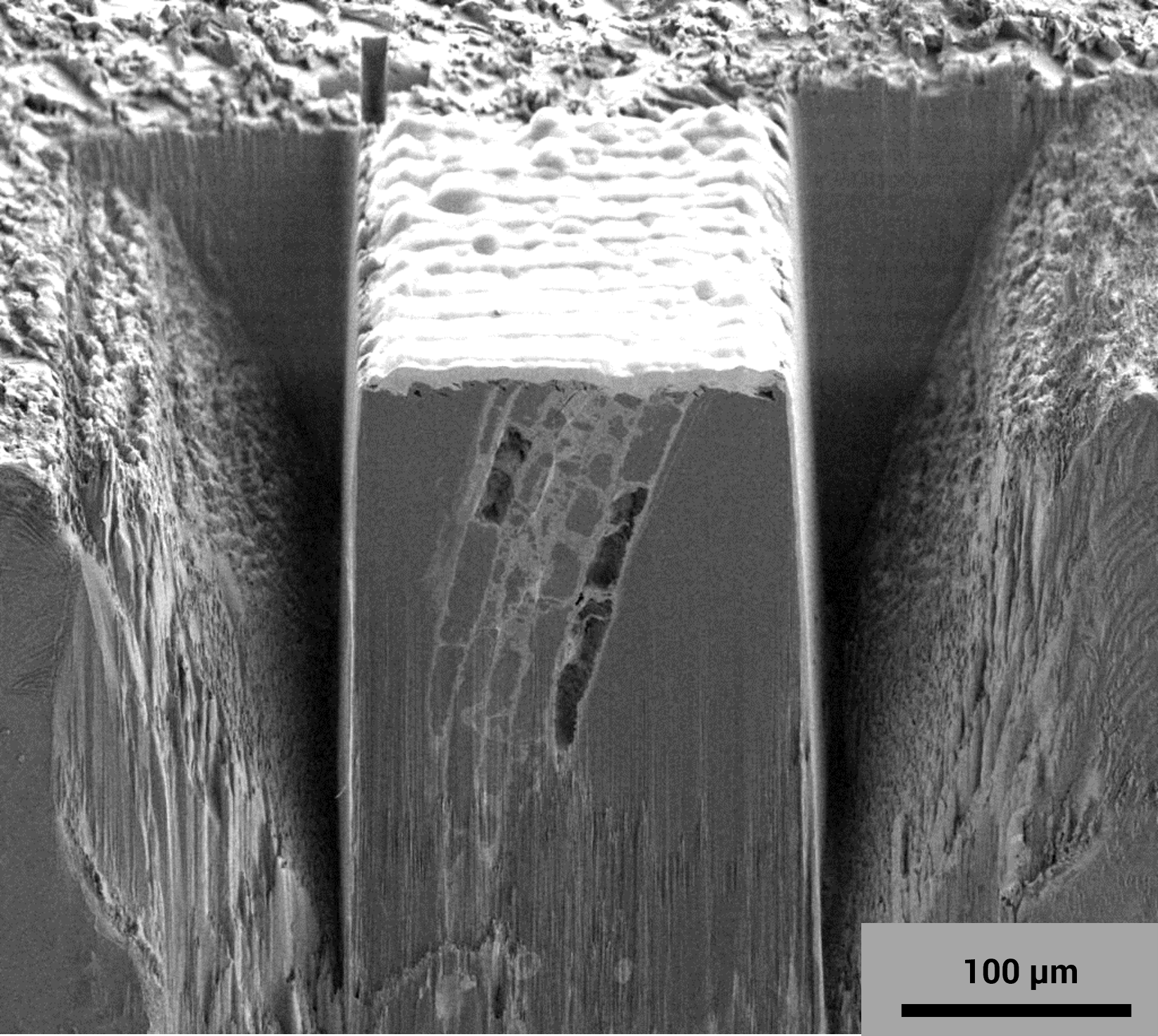
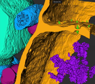
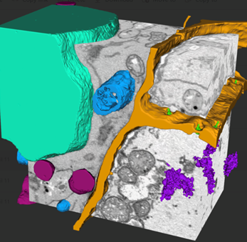
WEBINAR | Up to 40% Faster Automated TEM Lamella Preparation with new Ga⁺ FIB Column

Explore Intercellular Transport Pathways in Plants using Tescan Xe plasma FIB-SEM

Visualize intercellular transport pathways within plant tissue and uncover how nutrients move between cells through precise sub-micron 3D reconstruction using Tescan Xe plasma FIB-SEM.

4-1-Exploring Intercellular Transport Pathways in Plants
4-1-Exploring Intercellular Transport Pathways in Plants-mobile
Unlocked content

Study phloem structure in 3D with Tescan Xe plasma FIB-SEM

Intercellular transport in plants depends on delicate phloem structures like plasmodesmata in sieve cells. These structures are difficult to isolate and analyze using traditional imaging techniques.

TESCAN Xe Plasma FIB-SEM represents a powerful tool for Ultra-high resolution imaging and large-volume sample processing and reconstruction.
Study vascular architecture, trace transport pathways, and reconstruct phloem networks with the spatial detail to reveal how structure drives function.

How to Study Plant Intercellular Transport Pathways

with Tescan Xe Plasma FIB-SEM?

01
Root of the Problem

Why Conventional Imaging Fails to Capture fine Phloem Structures

Studying intercellular transport in plants depends on seeing the full 3D architecture of phloem tissue. But features like plasmodesmata are too small and deeply embedded for light microscopy or manual sectioning to capture reliably.

Focused Ion Beam (FIB) milling overcomes these limitations by sequentially removing ultra-thin slices of the sample. This achieves nanometer-scale resolution in the Z axis and enables accurate 3D reconstruction of fine phloem structures.

Tescan Xe Plasma FIB-SEM provides nanometer-scale resolution in the Z axis, enabling capturing tiny structures which would not be possible using an ultramicrotome.

Using FIB for milling provides superior Z resolution & 3D reconstruction of fine subcellular structures

Visualization and segmentation with Tescan Volume Analysis Software to underline transport pathways

Large-volume imaging preserves context for accurate biological interpretation

Explore how intercellular transport unfolds in plants using Tescan Xe Plasma FIB-SEM without losing spatial context or missing critical structural features.

02
Materials and Methods

How Plant Transport Structures Were Imaged Using Tescan Xe Plasma FIB-SEM

Resin-embedded tissue from Arabidopsis thaliana was trimmed to expose vascular regions containing phloem cells.

Automated FIB-SEM tomography was performed using Tescan Xe Plasma FIB-SEM, combining high-throughput Xe ion milling with Ultra-high resolution SEM imaging with in-beam Tescan Low-energy BSE detector at 2 keV. Each layer was sequentially milled away and imaged, producing ~900 of high-resolution images with 4 nm slice thickness and 4nm  pixel size.

3D volume reconstruction and segmentation were carried out using Tescan Volume Analysis Software. Researchers identified sieve cells, plasmodesmata, and internal organelles in 3D, with structural continuity across the tissue.

With this approach, researchers studied intercellular transport architecture in situ, preserving spatial relationships without distortion or loss of cellular context.

03
Results and Discussion

3D Visualization of Phloem Architecture in Plant Tissue

Tescan Xe Plasma FIB-SEM enabled researchers to acquire Ultra-high high resolution datasets from resin-embedded Arabidopsis phloem tissue. Serial sectioning with Xe plasma combined with Tescan Low-energy BSE detecor imaging revealed cellular structures which would be impossible to capture with conventional methods.

Sieve plates, cells, and plasmodesmata were clearly visualized in 3D, along with associated organelles and membrane interfaces. The reconstructed volumes revealed how phloem cells are spatially arranged and how structural changes such as cytoplasmic thinning and enucleation contribute to transport function.

With segmentation and 3D rendering, researchers identified transport pathways between cells and analyzed how architectural features influence molecular flow.

Sample courtesy of Dr. Ilya Belevich, EMBI, University of Helsinki.

GET IN Touch

Contact us

Get the most out of Tescan

This is more than information; it's an advantage. We've compiled our technical whitepapers, detailed product flyers, and on-demand webinars to provide you with the knowledge that makes a real difference. Sign up now to access the insights you need to make an impact.

Tescan Instruments & Technology

Used in This Workflow

Tescan SOLARIS X2

TESCAN Xe Plasma FIB-SEM enables high-resolution imaging, large-volume processing of resin-embedded plant tissue for structural analysis of phloem cells and transport pathways.

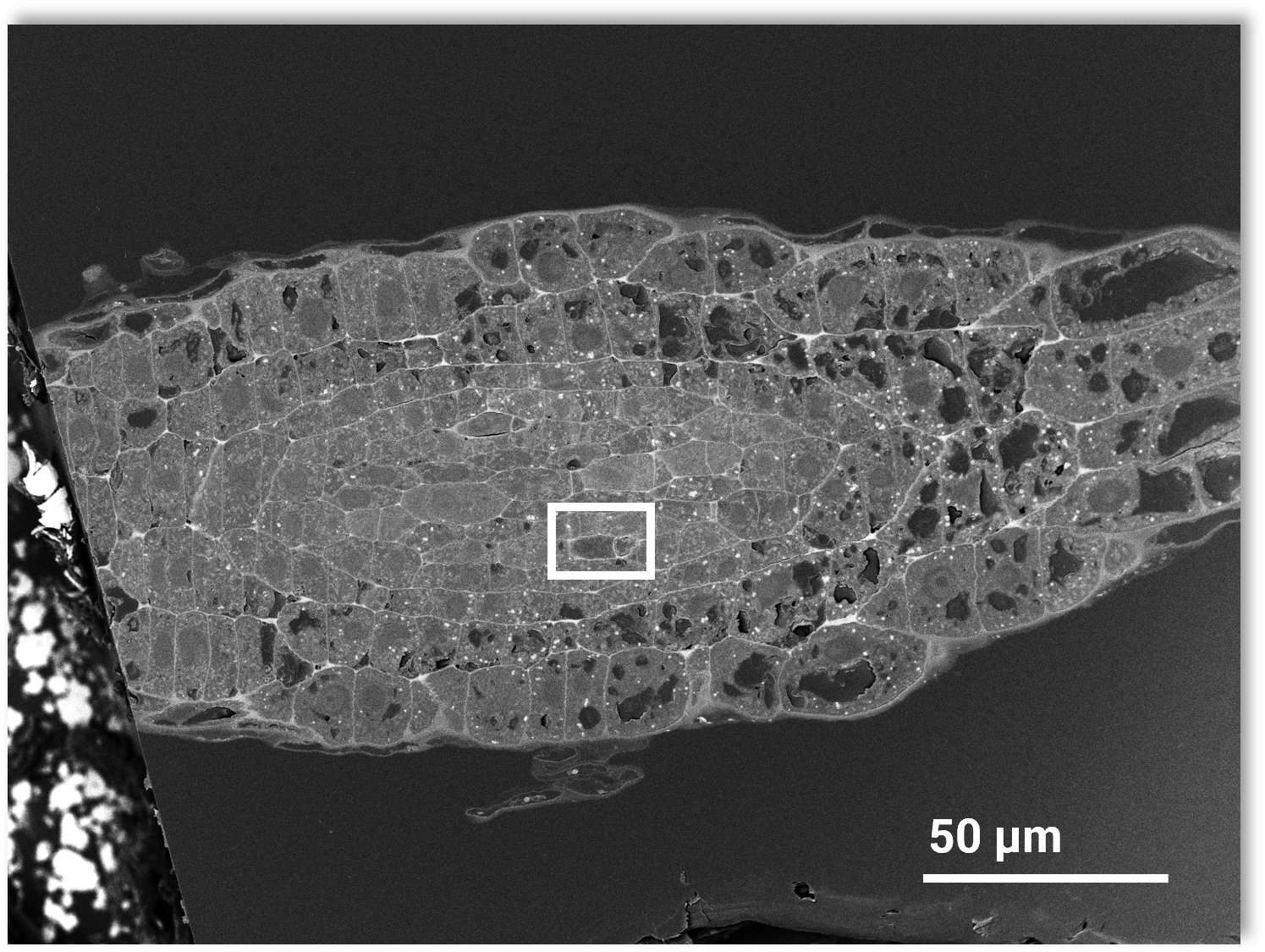
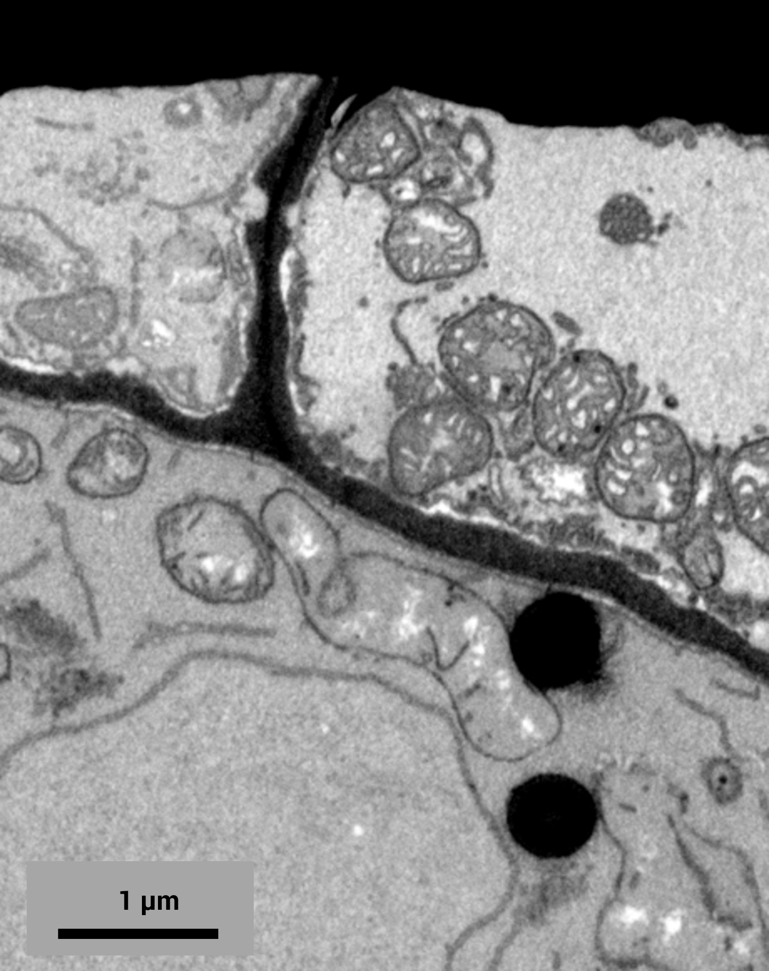
You can image sieve plates, plasmodesmata, and organelles in situ without manual sectioning, avoiding the compression and distortion associated with traditional slicing.

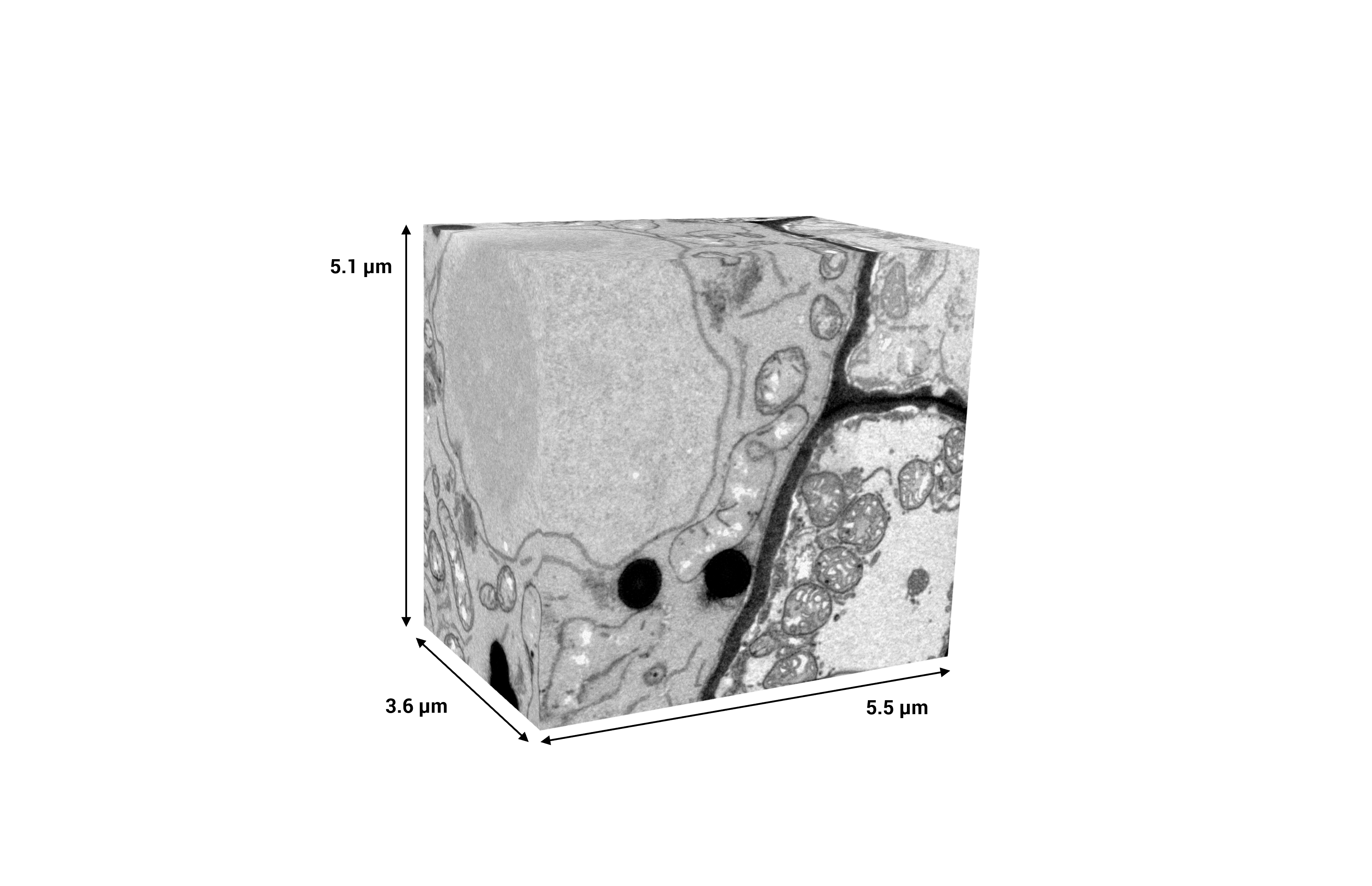
Wide-area plasma milling and automated acquisition give you the scale and precision needed to study delicate biological structures in full 3D.


  • MISTRAL Xe plasma FIB: allows you to remove material evenly without compressing or distorting biological tissue

  • Dedicated In-beam Tescan low-energy BSE detector: gives you high contrast for visualizing cellular boundaries and ultrastructure

  • Automated tomography workflow: lets you acquire consecutive slices with minimal setup or manual adjustment

  • 3D reconstruction across cell layers: helps you reveal full transport pathways and spatial cell organization

  • Tescan Volume Analysis Software: 3D vizualisation and segmentation

SOLARIS-X2 (2)

GET IN Touch

Contact us

map

Where can you find us:

Tescan Brno
Libušina třída 21
623 00 Brno
Czech Republic

info@Tescan.com