Micro-CT is a non-destructive X-ray imaging technique that creates detailed 3D models of an object's internal structure. In archaeology, it allows researchers to examine fragile artefacts, mummies, and ancient human remains without damaging the original specimen.
Listen to article
High-Resolution Tescan CoreTOM Imaging Reveals New Clues About an Iron Age Life and Death
Who Was Tollund Man?
Tollund Man, a remarkably preserved bog body discovered in Denmark in 1950, has fascinated researchers and the public for decades. Now, thanks to advancements in scanning technology, scientists are gaining unprecedented insights into this ancient individual.
How Micro-CT Revealed Hidden Details
In a recent secretive collaboration between Aarhus University and Museum Silkeborg, Tollund Man's head underwent a micro-CT scan using a Tescan CoreTOM system. This state-of-the-art technology offers up to 4000 times higher resolution compared to previous scans.
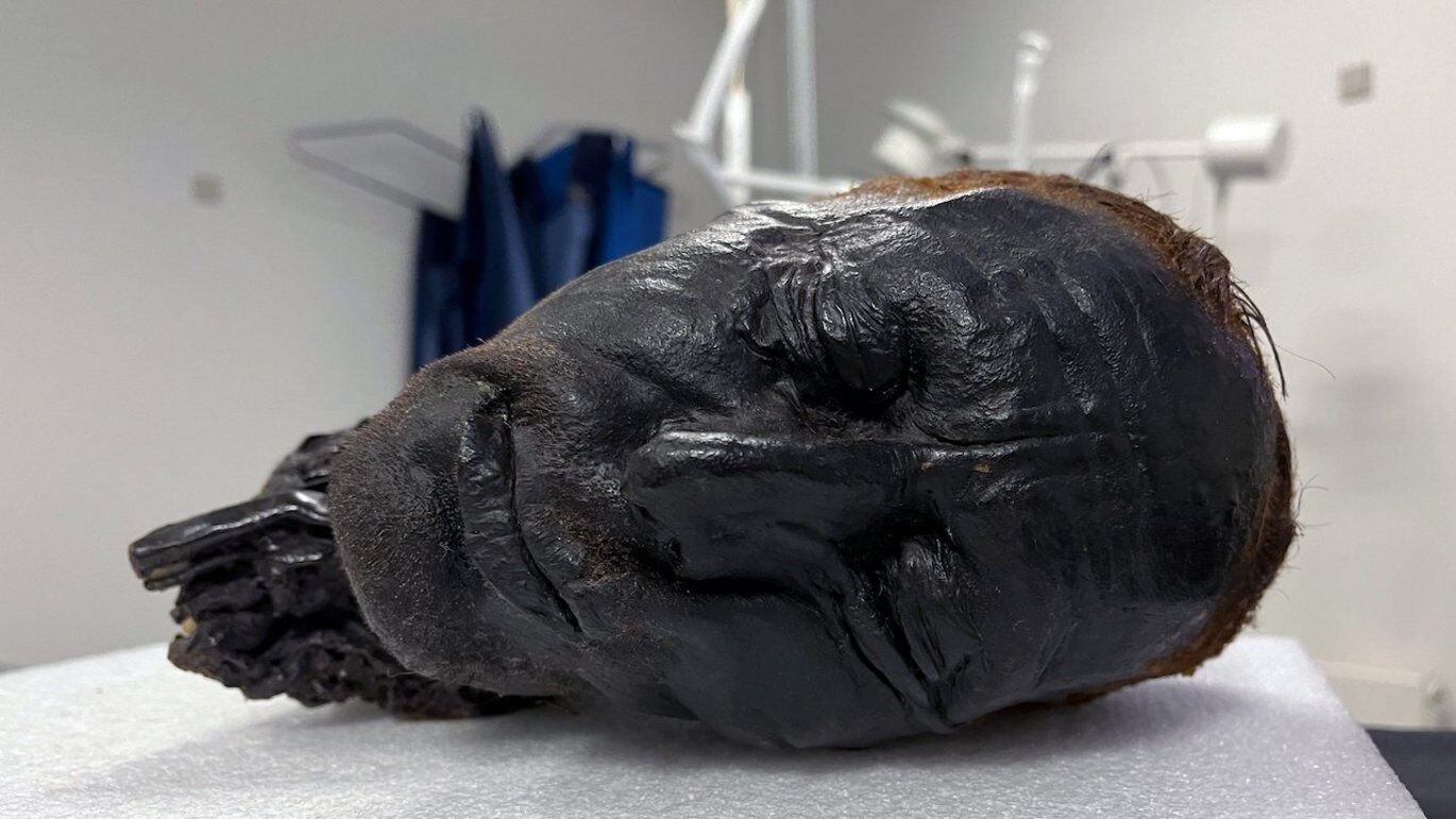 Figure: Tollund Man’s head is being prepared for the scan
The scan provided highly detailed images of the head's interior, including:
-
Teeth: Analysis of the teeth can reveal details about Tollund Man's diet, health, and age, potentially offering clues about his life experiences.
-
Eyes: The near-intact condition of one of Tollund Man's eyes allows researchers to study its shape and optic nerve, providing insights into his visual system.
Virtual autopsy of Tollund man's head and brain. With digital dissection, the brain can be viewed - it is well preserved.
Why Micro-CT Is Used in Archaeological Research
Micro-CT scanning, exemplified by the Tescan CoreTOM system, is versatile with applications beyond forensic investigations. Researchers can analyze:
-
Clinical and forensic tissue samples for medical insights.
-
Specimens of laboratory animals to study treatment effects.
-
Soil samples to understand composition and structure.
-
Museum objects non-destructively, revealing hidden details and aiding in preservation.


How Tescan CoreTOM Supports Archaeological Imaging
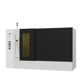
The Tescan CoreTOM Multi-resolution micro-CT system, offers vast capabilities:
-
Multi-scale Imaging: It can image full cores up to 1 meter in length and millimeter-sized samples, allowing for both broad and detailed examination.
-
High Throughput: Optimized for fast scans, it maximizes efficiency, enabling more sample analysis.
-
Volume of Interest Scanning (VOIS): Researchers can focus high-resolution scans on specific critical areas.
-
In Situ Integration and Dynamic CT: The CoreTOM allows for external device integration for in situ experimentation.
-
Flexibility for Research: Tescan offers open access to scripting protocols and raw data, empowering deeper research exploration.
What the Scans Revealed About Tollund Man
The results of the Tollund Man scan using the Tescan CoreTOM are still being analyzed but promise to enhance our understanding of this Iron Age individual. By studying his teeth, eyes, and other features, researchers may better determine his cause of death, reconstruct his diet and health, and gain insights into his social status.
Tollund Man's story highlights the power of bog preservation and the ongoing quest to unlock the secrets of the past. With advancements like micro-CT scanning technology, particularly systems like the Tescan CoreTOM, researchers can now delve deeper than ever before, bringing the lives of our ancestors into sharper focus.


Explore Micro-CT Resources
UniTOM® XL
Time-resolved micro-CT for dynamic process imaging.

Geo Sciences
Explore micro-CT for geoscience and cultural heritage

Discuss Your Sample Preparation Workflow with Our Team