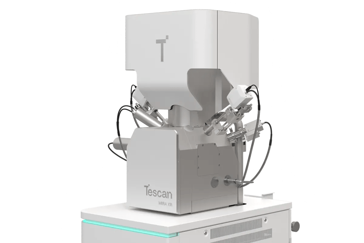
WEBINAR | From Infrastructure to Impact: EM & Micro-CT in Materials Core Facilities

Morphology of Mesoporous Silica Revealed by Low-kV SEM Imaging

Tescan MIRA XR provides high-resolution insight into SBA-15 particle shape and structure.

hero
hero
Unlocked content

From Macroscale Particle Size Determination to Nanoscale Pore Structure in One Workflow

Mesoporous silica materials like SBA-15 are valued for their well-ordered pores, large surface area, and tunable pore size. But to evaluate them properly, especially in their native form, the imaging method needs to match the material’s sensitivity. Too much energy, and delicate details are lost.

This study demonstrates how Tescan MIRA XR, equipped for low-voltage, high-resolution SEM imaging, allows researchers to directly observe nanorod morphology and porosity in SBA-15 without charging, coating, or damage. This is especially beneficial when performing investigations of beam-sensitive nanoscale materials.

What Makes

This Imaging Approach Different?

01
Root of the Problem

Gentle Imaging for Fragile Structures

Porous nanoparticles like SBA-15 are inherently sensitive. Their usefulness in catalysis, drug delivery, and molecular separation depends on pore structure and size, and the resulting surface area. Conventional SEM can damage the uncoated surfaces, and other analytical techniques may not be able to accurately capture surface detail.

Transmission electron microscopy (TEM) offers excellent resolution but often lacks context and requires thinning. Standard SEM, meanwhile, can produce charging artifacts unless the sample is coated. The challenge was to find a way to see SBA-15’s real surface structure without altering it. 

02
Materials and Methods

Low-Voltage SEM Imaging of As-Synthesized Mesoporous Powders

The sample was as-synthesized mesoporous silica SBA-15 powder, produced using tetraethoxysilane (TEOS) and a triblock copolymer template. The powder was mounted directly onto a conductive carbon tape without additional coating or processing.

Imaging was performed on a Tescan MIRA XR using BrightBeam™ optics in low-voltage conditions: 800 eV acceleration voltage, 10 pA beam current, and an in-column Axial detector, with each field of view acquired in under one minute—imaging parameters that reduce charging and preserve the synthesized sample.

The resulting images showed both rod-like and curved particle shapes, along with nanoscale pore structure, providing a basis for qualitative and statistical shape assessment.

03
Results and Discussion

Native Morphology, Clearly Captured

SEM imaging under these conditions produced high-contrast views of individual SBA-15 particles. Surface porosity, curvature, and length variation were recorded with each image acquisition, delivering sufficient data to support meaningful statistical analysis of the particles.

Notably, no conductive coating was used, and the applied beam parameters suppressed charging artifacts. This indicates that low-voltage SEM imaging, when properly configured, is sufficient for direct analysis of non-conductive nanostructures like mesoporous silica.

Tescan MIRA XR provides a practical tool for labs investigating nanoscale materials properties, connecting micro- and nanoscale structure to synthesis effects, scaling consistency, or application-specific morphology in porous nanomaterials.

GET IN Touch

Contact us

Get the most out of Tescan

This is more than information; it's an advantage. We've compiled our technical whitepapers, detailed product flyers, and on-demand webinars to provide you with the knowledge that makes a real difference. Sign up now to access the insights you need to make an impact.

Tescan Instruments & Technology

Used in This Workflow

Tescan MIRA XR

A versatile UHR-SEM system for beam-sensitive samples.

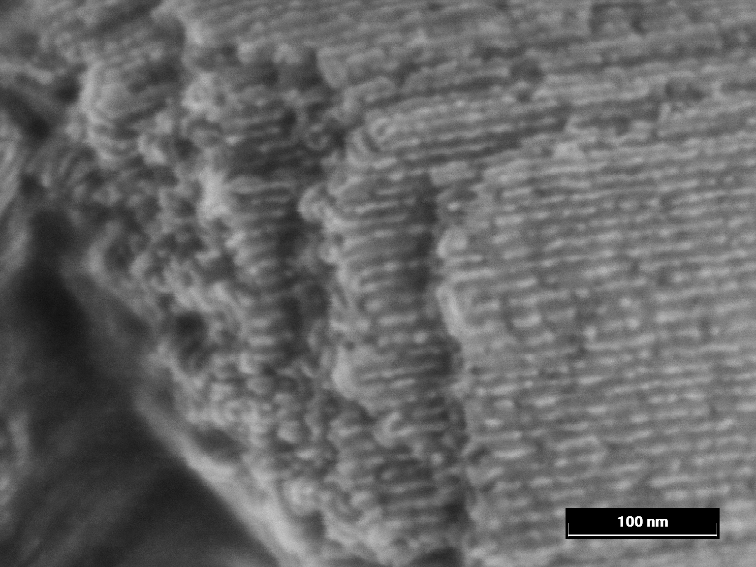
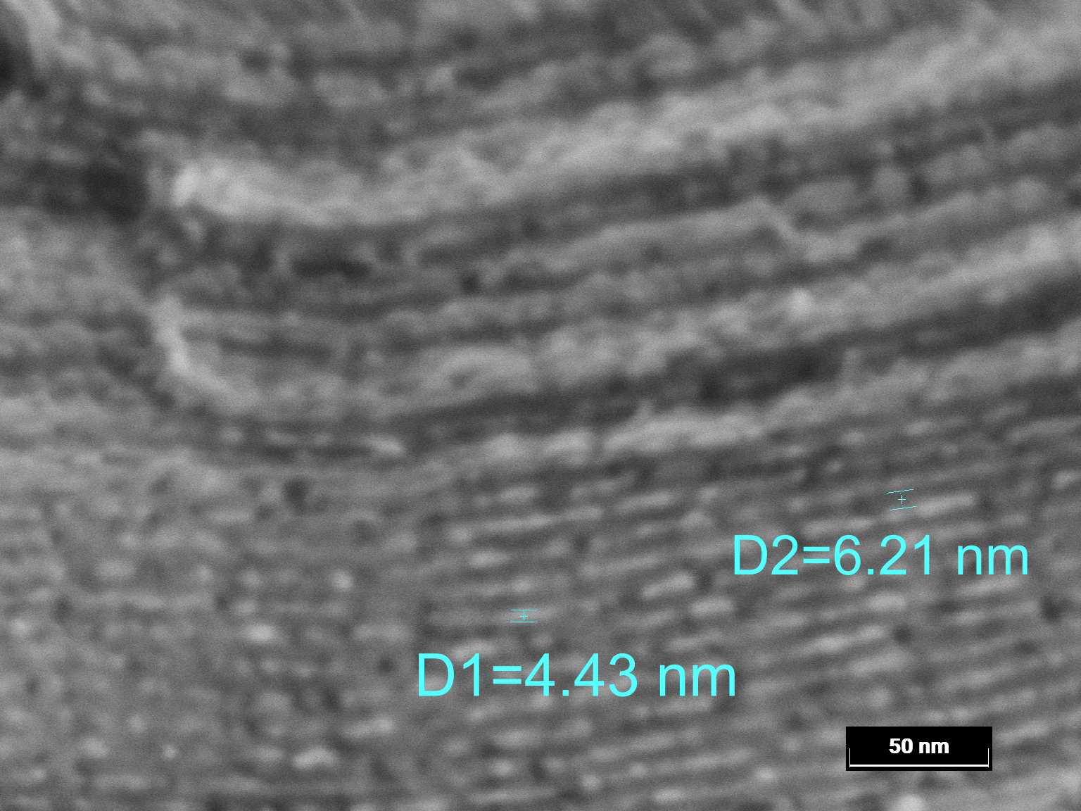
  • Reveal fine surface details with BrightBeam™ electron optics and efficient signal collection

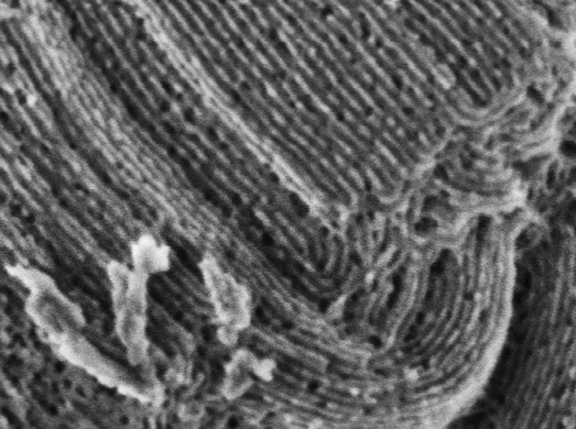
  • Preserve delicate or uncoated samples through low-voltage imaging

  • Achieve sub-nanometer resolution with optimized in-column detection for nanoscale surface information

MIRA XR GM MONO Metal

GET IN Touch

Contact us

map

Where can you find us:

Tescan Brno
Libušina třída 21
623 00 Brno
Czech Republic

info@Tescan.com 

130405923 us US 37.09024 -95.712891 25.3575 29.349345 20.67957527 42.082797 39.91384763 -33.693421 13.93320106 3.039986586 31.997988 38.050985 47.579533 48.1485965 58.375799 54.663142 19.195447 56.975106 50.493053 45.868592 10.79556993 44.35660598 43.2371604 55.536415 14.557577179752773 32.100937 -6.116829 -6.212299277967318 23.7104 -33.471062 31.998740087 -23.69149395 43.462349 51.529848 49.1893523 49.197486 25.072375 31.075811 1.299027 40.676979 52.30150662 51.013813 35.684121 37.479653 52.246622 40.581349 39.911632 -26.1811371 41.818215 33.429928

No distributors found.