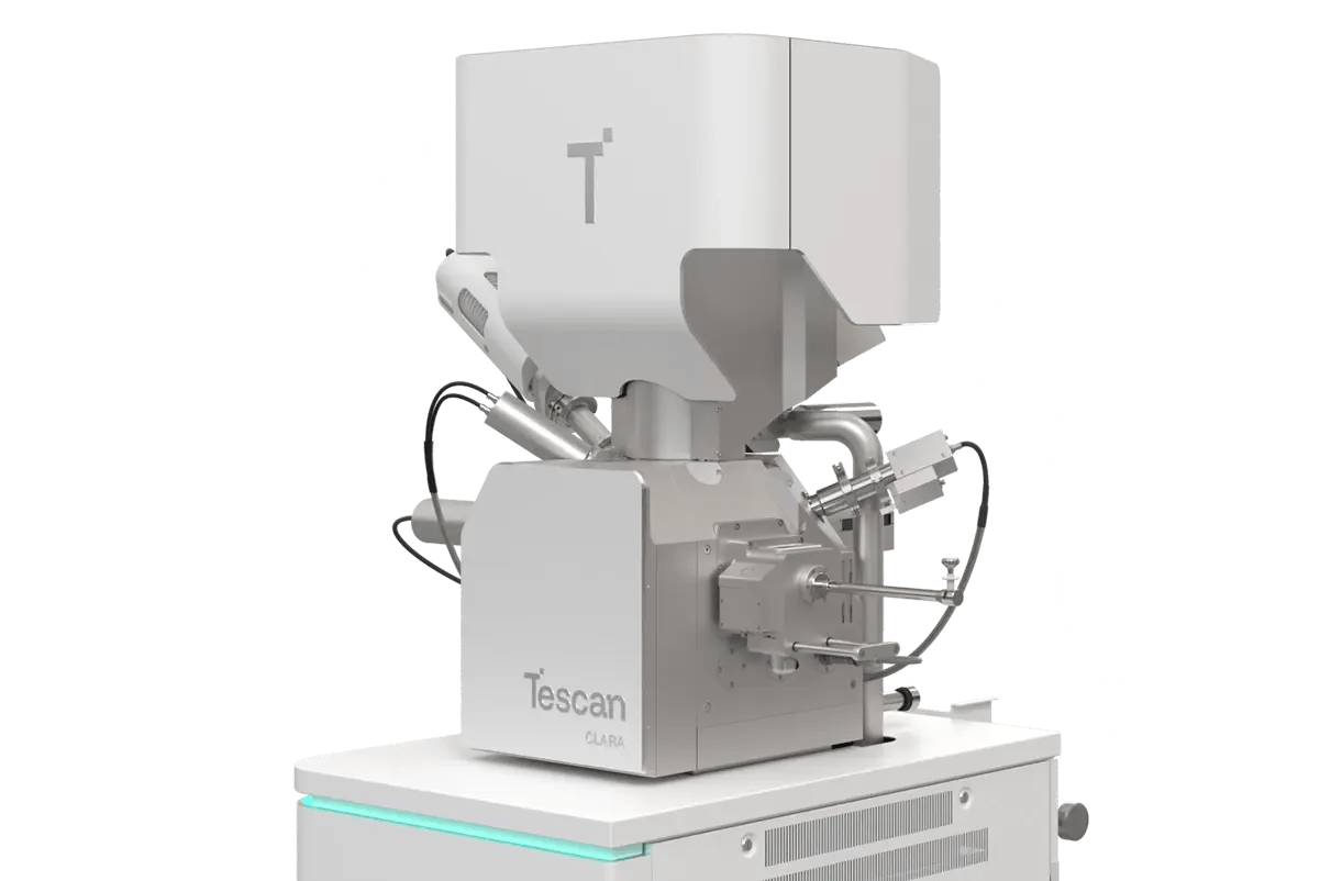
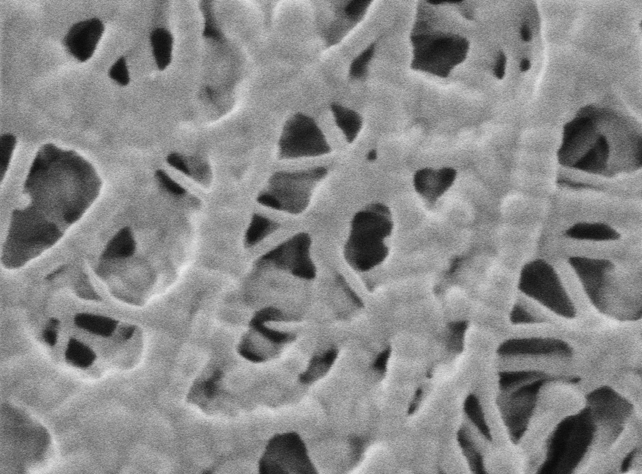
WEBINAR | Up to 40% Faster Automated TEM Lamella Preparation with new Ga⁺ FIB Column

Low-kV SEM Imaging of Separator Porosity in Lithium-Ion Batteries

Tescan CLARA delivers pore-level insight into beam-sensitive polymer membranes.

hero
hero
Unlocked content

Characterizing What Matters

Battery separators are often overlooked in the broader cell architecture, but they carry significant influence over safety and ionic conductivity. Their fibrous, porous design must strike a balance: open enough to allow ion flow, but strong enough to maintain structural separation between electrodes.

To evaluate this balance, researchers used Tescan CLARA’s ultra-high-resolution SEM to image a commercial polypropylene separator without surface coating. Operating at just 200 eV, the system revealed clear pore networks and fiber architecture without damaging the material, offering direct access to structural metrics critical to separator performance.

Why Use Low-kV SEM

for Separator Imaging?

01
Root of the Problem

Seeing Pores Without Obscuring Them

Battery separators are built to manage ion flow while physically isolating electrodes. This dual role depends on pore structure; size, shape, and distribution all play into ionic transport and mechanical resilience. But imaging these details is not straightforward.

Standard SEM imaging of separators often requires metal coating, which can obscure fine pore dimensions. Alternatively, imaging uncoated samples at low beam energies and currents leads to resolution loss in conventional SEMs. This study demonstrates how ultra-high-resolution SEM enables detailed pore structure analysis without compromising resolution or sample integrity. 

02
Materials and Methods

The sample was a commercially available polypropylene (PP) separator, analyzed as received, with no conductive treatment. Imaging was performed with the Tescan CLARA, at 200 eV and using an axial detector specifically chosen for high-contrast topographic response.

Key imaging conditions:

  • No coating

  • Beam energy: 200 eV

  • Magnifications tuned for both pore network overview and fiber detail

Captured images were processed using MIPAR software; automated segmentation tools were applied to:

  • Measure pore diameters (range: 20 to 140 nm; average: 59 nm)

  • Evaluate fiber thickness (range: 20 to 211 nm; average: 71 nm)

03
Results and Discussion

Pore Clarity Without Sample Prep

Even at this low beam energy, the SEM produced crisp images of the membrane surface. Pore edges were well-defined, allowing segmentation and measurement without manual tracing or correction. Fiber width and pore spacing showed significant heterogeneity, emphasizing the importance of direct analysis over assumptions based on material formulation.

The ability to image uncoated polymer samples at nanometer resolution opens new possibilities for separator quality control and materials development, especially when porosity must be matched to electrolyte viscosity or thermal shutdown behavior.

With this method, users gain access to fine-scale features without sacrificing time or damaging delicate samples.

GET IN Touch

Contact us

Get the most out of Tescan

This is more than information; it's an advantage. We've compiled our technical whitepapers, detailed product flyers, and on-demand webinars to provide you with the knowledge that makes a real difference. Sign up now to access the insights you need to make an impact.

Tescan Instruments & Technology

Used in This Workflow

Tescan CLARA

A precision SEM platform designed for low voltage operation and exceptional surface contrast.

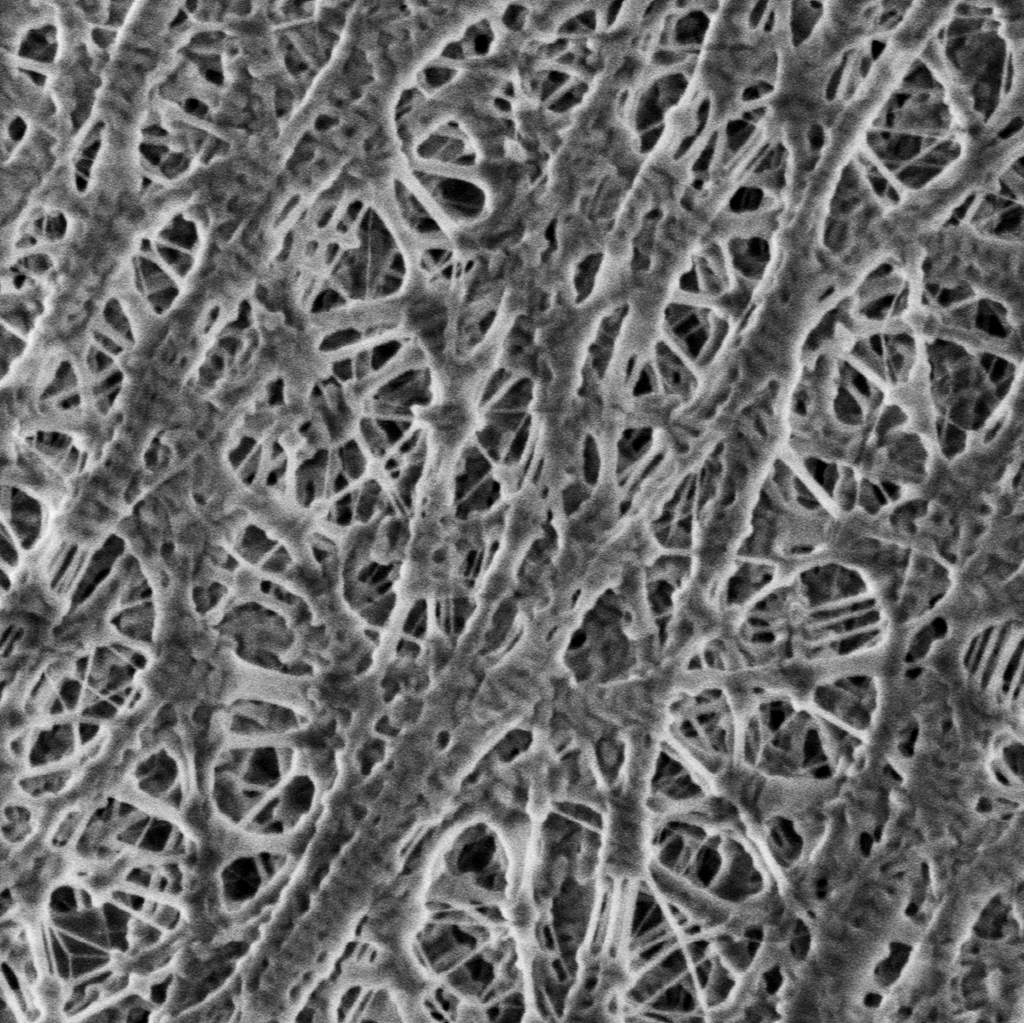
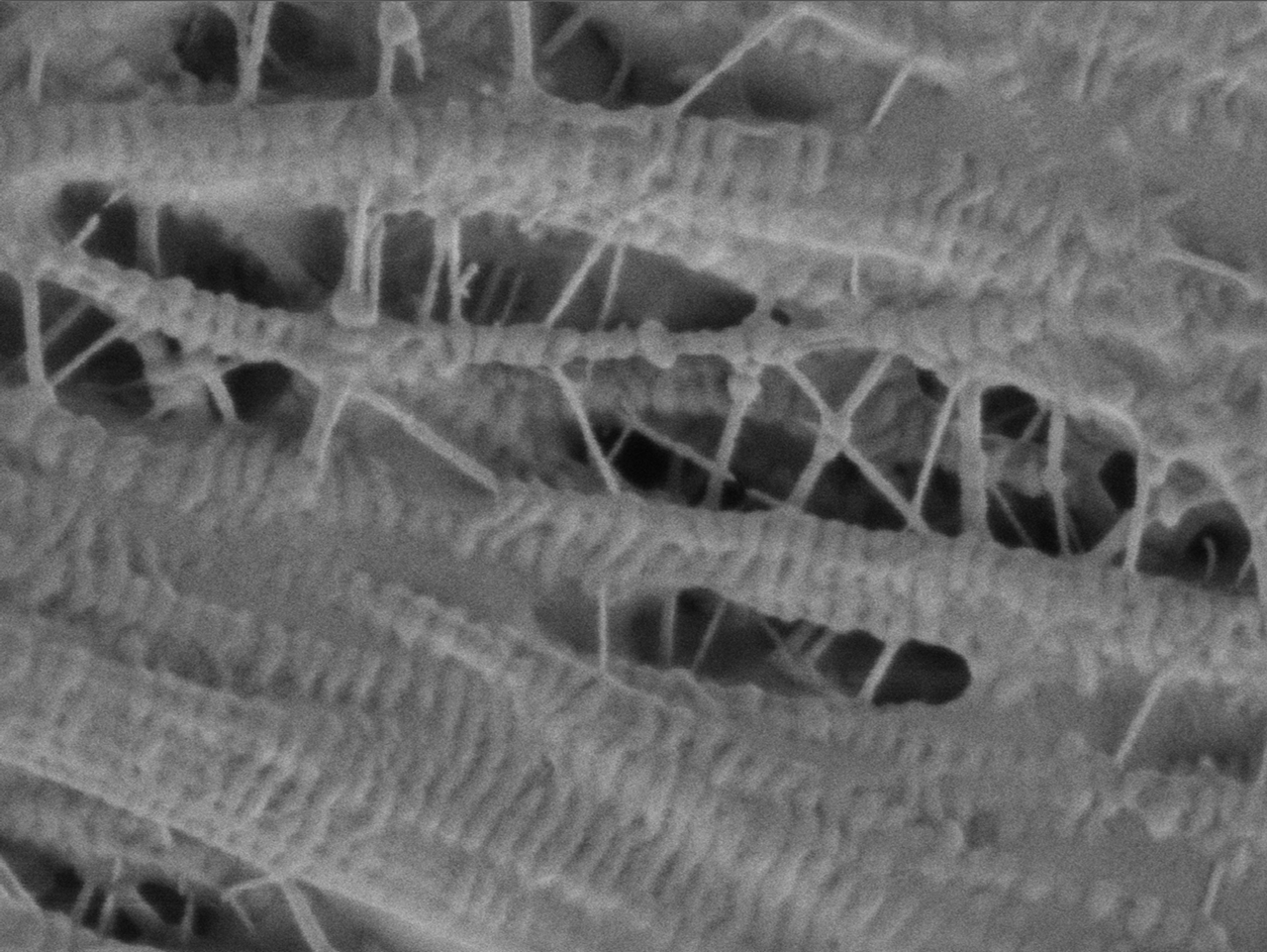
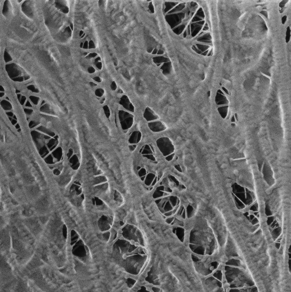
  • Performs at low voltages to preserve beam-sensitive materials

  • Delivers clear imaging of uncoated polymers and composites

  • Ideal for porous membranes, fibrous films, and soft nanostructures

CLARA GM Mat. Science

GET IN Touch

Contact us

map

Where can you find us:

Tescan Brno
Libušina třída 21
623 00 Brno
Czech Republic

info@Tescan.com