Achieve Consistent Results
with Electron Microscopy that

Supports your Next
Breakthrough

Accelerate your research with microscopy tools that simplify workflows and deliver consistent outcomes to support your next breakthrough.

Bringing added value across industries

with Electron Microscopy Solutions
_ Application area icon
Material Science
Optimize Material Performance with High-Resolution Imaging

Develop stronger, lighter, and more durable materials with Tescan’s SEM and FIB-SEM solutions. Gain precise structural and compositional insights for failure analysis, quality control, and material innovation.

  • 3D characterization
  • Non-destructive 3D analysis
  • Surface analysis
  • Nanoprototyping
  • TEM sample preparation
Material_Science_1x-1
_ Application area icon (1)
Semiconductors
Pinpoint Defects and Root Causes Using High-Precision FIB-SEM Tools

Streamline development and quality control with Tescan’s FIB-SEM and STEM systems. Gain accurate, nanoscale insight into structures, interfaces, and defects to optimize fabrication processes and extend device performance.

  • Defect localization and failure analysis
  • Delayering and cross-sectioning
  • Root cause analysis
  • Circuit modification and nanoprototyping
  • TEM lamella preparation
pic1_1x
_ Application area icon (4)
Batteries
Study Sensitive Battery Interfaces with Low-Damage, High-Resolution Imaging

Reveal electrode structure, monitor interface degradation, and detect dendrite growth using Tescan’s SEM, FIB-SEM, and cryo workflows. Preserve fragile chemistries while gaining structural and compositional insight across ambient and cryogenic conditions.

  • Lithium dendrite detection
  • Electrode–electrolyte interface analysis
  • Cryo-FIB sample preparation
  • In situ and 3D imaging workflows
  • Structural and chemical degradation studies
Batteries_1x
_ Application area icon (1)
Life Science
Achieve Structural Clarity and Preserve Biological Integrity with Cryo-FIB-SEM

Visualize ultrastructure across cells, tissues, and organoids with imaging workflows designed to protect delicate biological samples. Tescan’s Cryo-FIB-SEM enables high-contrast imaging, precise sample preparation, and consistent results — all while preserving native structure.

  • Cryo-FIB-SEM imaging
  • Correlative light and electron microscopy (CLEM)
  • Volume EM and 3D reconstruction
  • Organoid and tissue-level analysis
Life_Science_1x
_ Application area icon
Geoscience
Visualize Rock Microstructure and Composition in 3D with FIB-SEM and Micro-CT

Analyze rock cores, cuttings, and thin sections with workflows that reveal pore morphology, mineral composition, and structural variation. Tescan’s FIB-SEM and micro-CT systems support non-destructive 2D and 3D imaging for high-resolution insight across geological samples.

  • Mineral mapping and compositional contrast
  • Pore network and permeability analysis
  • 3D petrography and core characterization
  • Automated mineralogy and particle analysis
  • Quantitative EDS and BSE workflows
Geoscience_1x

Tescan Instruments

for every electron microscopy 
workflow
Tescan’s portfolio spans SEM, FIB-SEM, 4D-STEM, and microCT systems—designed to serve advanced research, development, and quality control. From nanofabrication and TEM sample prep to non-destructive 3D imaging, our instruments combine precision, flexibility, and application depth across every major scientific and industrial domain.

Whether you're solving challenges in failure analysis, technology pathfinding, or process optimization, Tescan systems adapt to your goals—today and in the future.

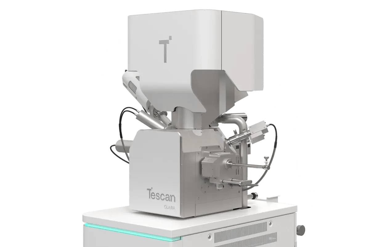
Scanning Electron Microscopes (SEM)

Tescan’s SEM platforms provide high-resolution imaging and surface analysis with exceptional contrast, detail, and ease of use—ideal for materials research, quality control, and analytical applications.

CLARA GM Mat. Science

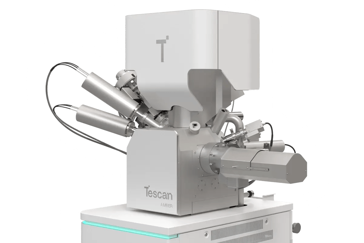
Focused Ion Beam-Scanning Electron Microscopes (FIB-SEM)

Tescan FIB-SEM systems combine fast, precise milling with high-resolution imaging and advanced automation. Choose from Ga or Xe plasma FIB sources to match your needs in sample prep, prototyping, or failure analysis.

AMBER 2

Micro-computed tomography (microCT)

Tescan’s X-ray micro-CT systems deliver fast, high-resolution 3D and 4D imaging for non-destructive internal analysis. Dynamic in-situ capabilities and modular designs make them ideal for advanced research across materials, life sciences, and energy storage.

MICRO_UniTOM_HR_1-2

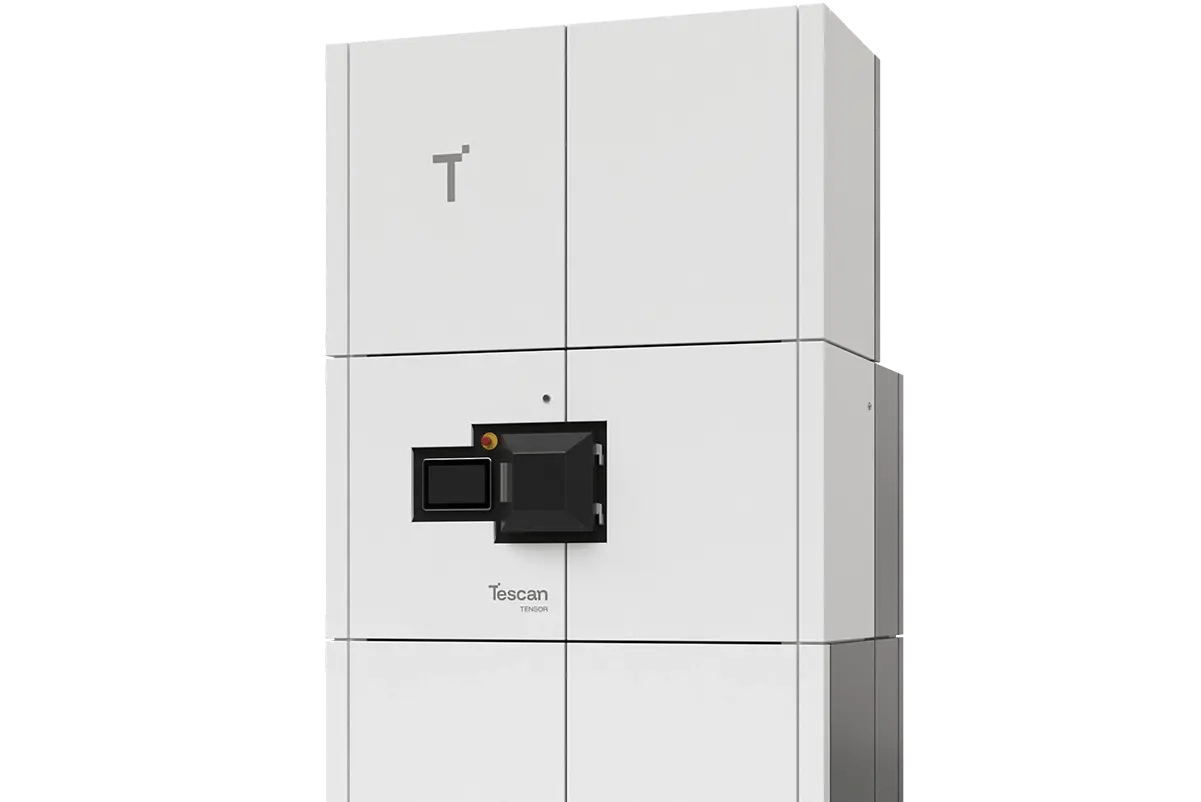
4D STEM

Tescan's TENSOR is the first 4D-STEM platform built for intuitive multimodal nanocharacterization, combining structural, morphological, and chemical insights in every scan. Real-time data processing, automation, and scripting support make it ideal for both routine and advanced research.

TENSOR_1