Image Complete Batteries Without Sectioning
Perform non-destructive 3D imaging of full pouch and prismatic cells to study internal architecture, interfaces, and assembly precision across multiple length scales.

Advance battery inspection and in situ analysis with the Tescan UniTOM® XL. Designed for large-scale micro-CT, it enables multiscale imaging of pouch and prismatic cells, 4D dynamic CT of degradation processes, and real-time visualization of structural changes during cycling.
.png?width=1890&height=1260&name=5_vertual%20slice%20through%20pouch%20cell%20highlighting%20cracks%20(1).png)
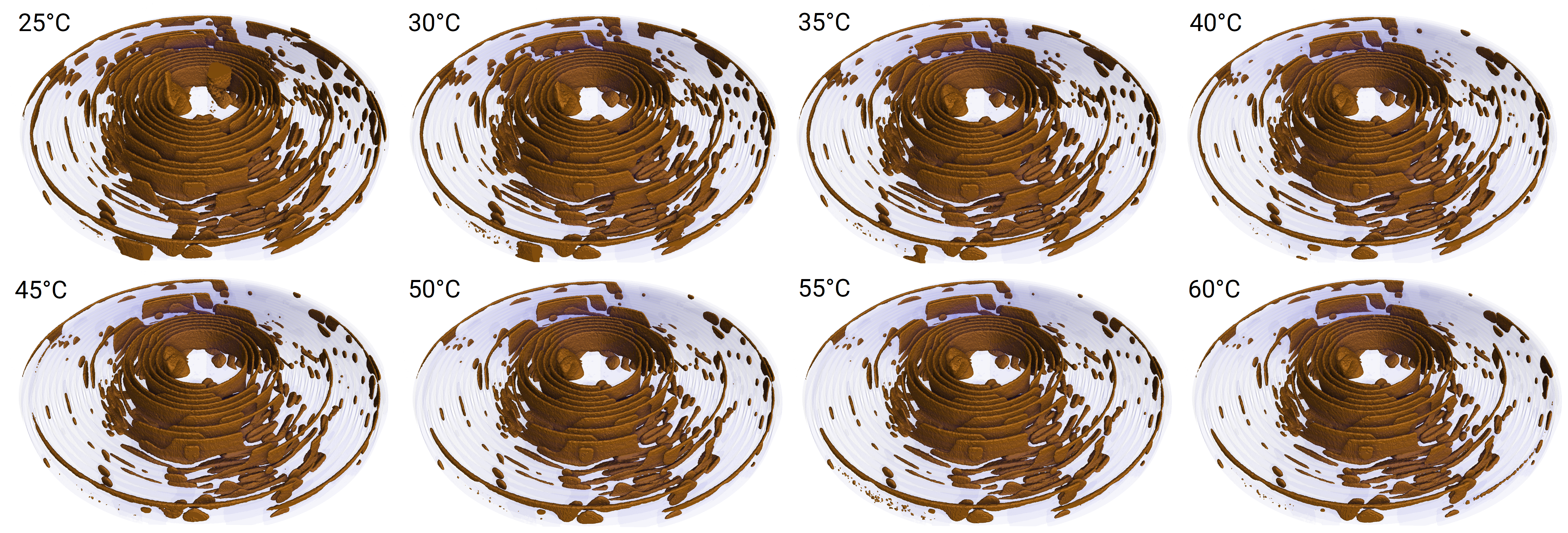
Track thermal effects, gas evolution, and electrolyte movement in lithium-ion battery heating using non-destructive 4D imaging with Tescan UniTOM® XL.
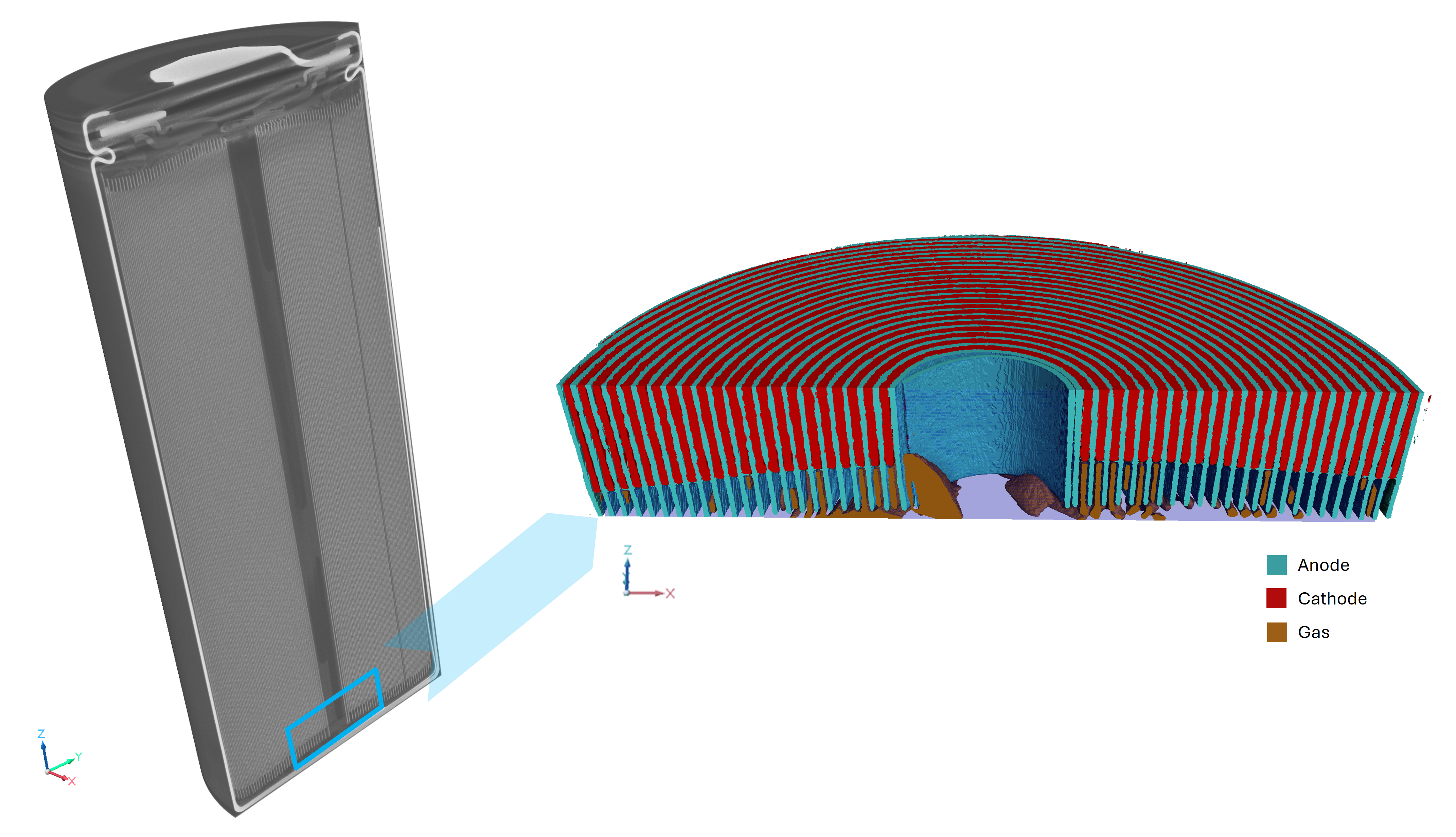
.png?width=1547&height=1161&name=3_Detailed%20micro-CT%20analysis%20of%20anode%20overhang%20in%20a%20cylindrical%20battery%20cell%20(1).png)
High-resolution X-ray tomography uncovers subtle but critical anode overhang variations — helping battery developers align performance with safety standards.
%20(1).webp?width=1698&height=1199&name=5_Non-destructive%20analysis%20of%20anode%20overhang%20in%20a%20cylindrical%20battery%20cell%20(1)%20(1).webp)
X-ray microtomography provides a non-destructive view into internal cell architecture — supporting performance validation, design optimization, and safety-critical QA.
%20(1).webp?width=470&height=456&name=Nov%C3%BD%20projekt%20(7)%20(1).webp)
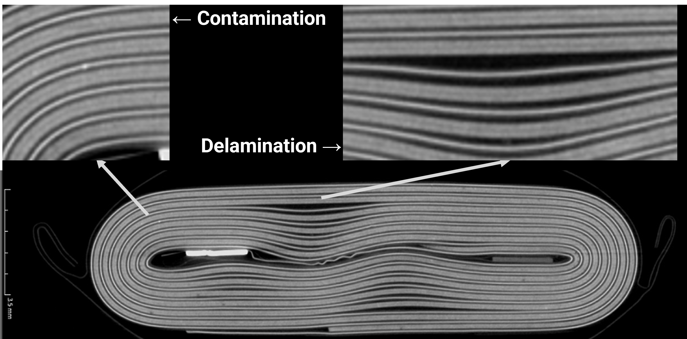
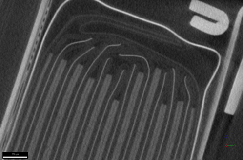
Detect delamination and material inconsistencies inside compact wearable battery systems using high‑resolution spectral micro‑CT for battery analysis – non‑destructively and in full 3D.
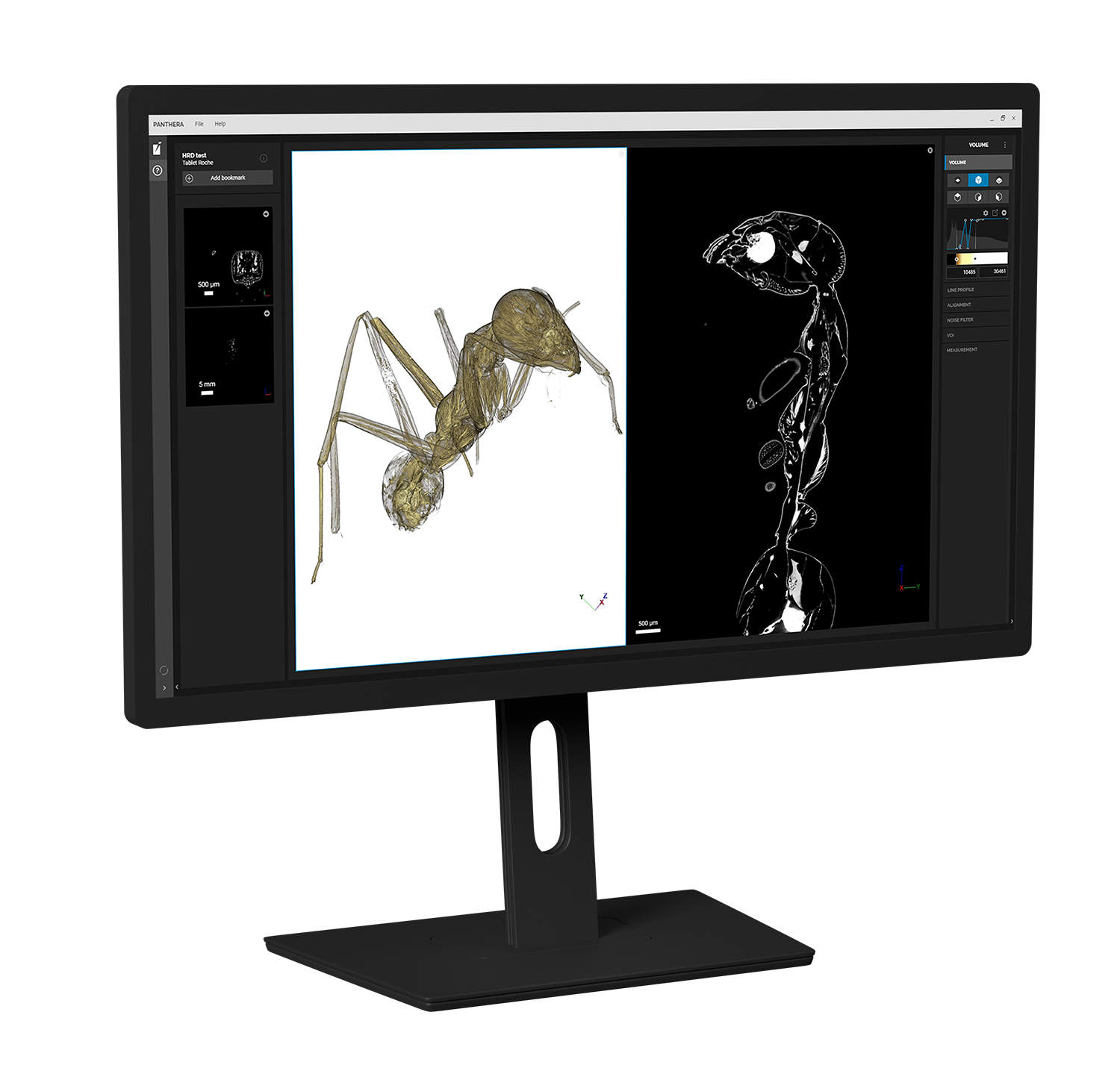
Panthera™ delivers GPU-optimized reconstruction and visualization of large 3D and 4D datasets.
Batch reconstruction and Volume of Interest Selection (VOIS) streamline multiscale workflows, while integrated filtering and temporal alignment ensure consistent data quality. From high-throughput QC to in situ analysis, Panthera™ provides the speed and accuracy needed for dependable imaging results.
TEM AutoPrep™ Pro automates the complete TEM lamella preparation process, delivering clean, Ga-free specimens optimized for nanoscale battery analysis. Integrated into the Essence™ GUI, AutoPrep™ Pro removes operator variability and ensures reproducibility across multiple samples and sites.
AI-guided routines handle trench milling, lift-out, grid placement, and 500 eV final polishing, producing lamellae with the right thickness and orientation for high-resolution STEM, EDS, and ToF-SIMS correlation. Multi-site batch operation enables unattended overnight preparation, saving valuable operator time.
With TEM AutoPrep™ Pro, battery researchers and manufacturers can depend on consistent, high-quality lamellae that reduce preparation time and accelerate the investigation of critical interfaces and degradation mechanisms.
.jpg?hsLang=en)


.webp?hsLang=en)