

.webp?hsLang=en)
.webp?hsLang=en)
Tescan UniTOM® XL
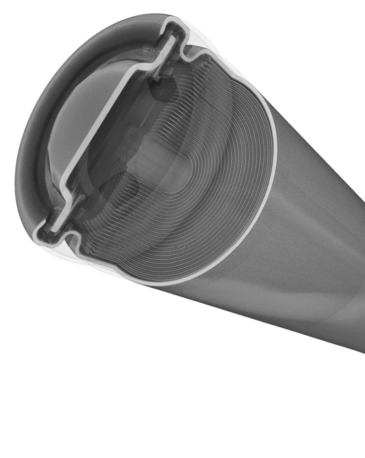
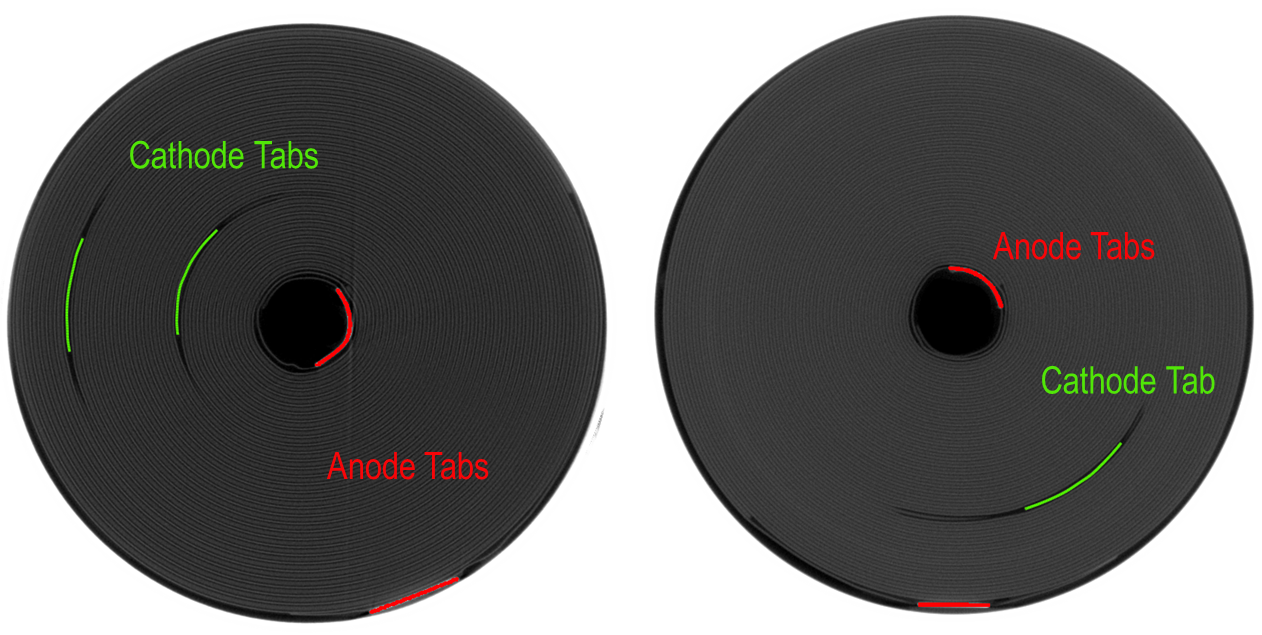
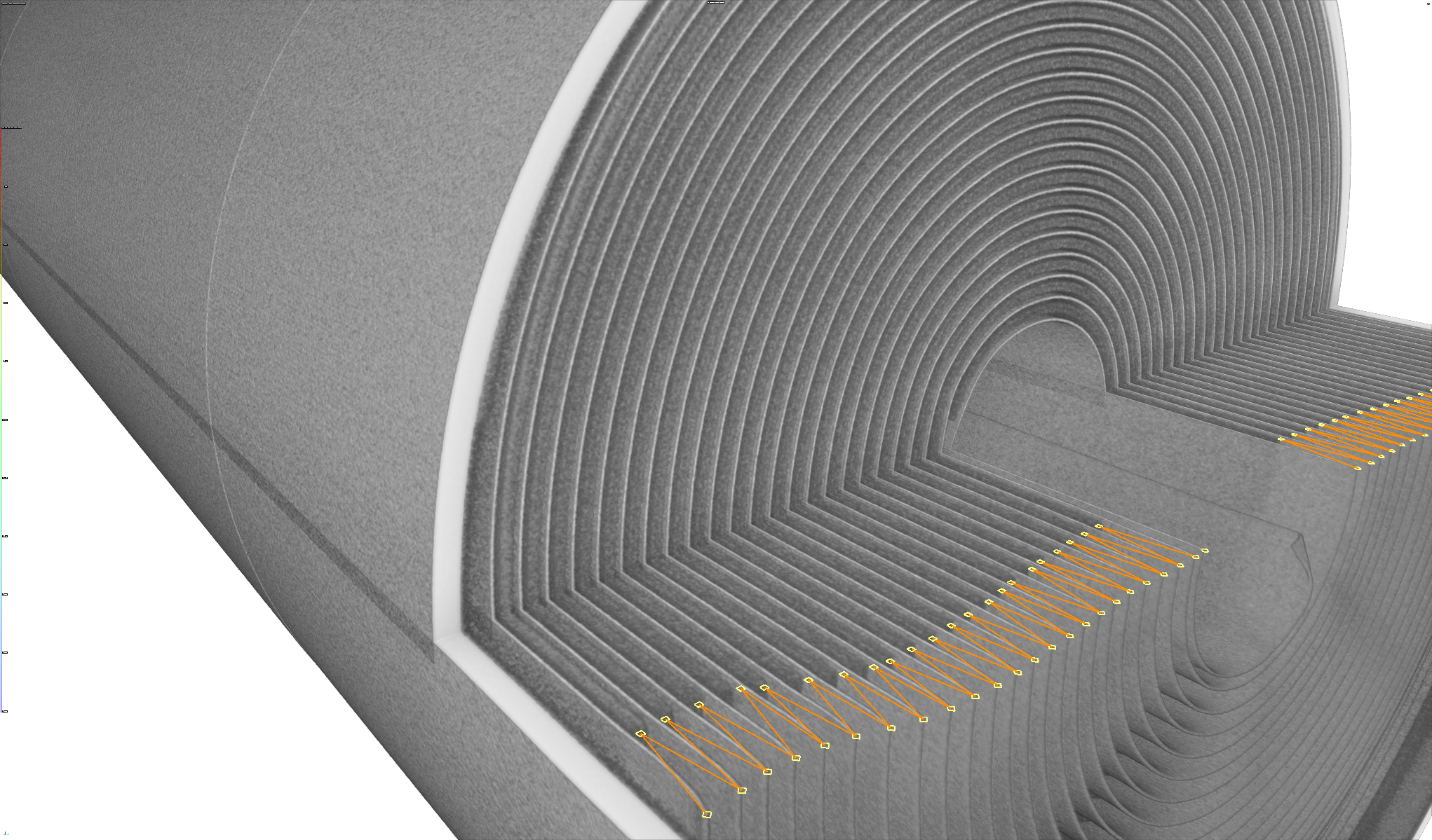
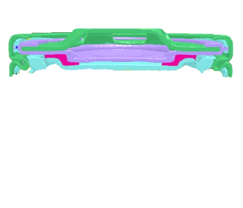
Tescan UniTOM® XL combines speed, resolution, and large sample volume capacity, enabling complete, non-invasive imaging of cylindrical, prismatic, and pouch cells.
- Supporting high-resolution scans of full battery cells and modules
- Fast reconstruction of internal components with no sectioning required
- Suitable for inspection of tab structure, cap assembly, and layer alignment
- Seamless workflow with segmentation and analysis in VGSTUDIO MAX


