Capture Nanoscale Surface Detail with Confidence
Capture nanoscale surface detail with confidence. BrightBeam™ optics reveal the intricate features of battery materials with exceptional clarity and resolution.
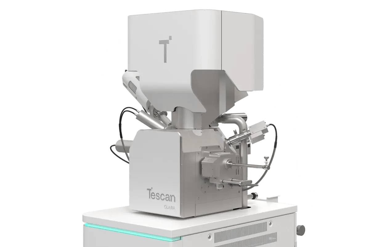
Achieve nanoscale precision in imaging and analysis with Tescan CLARA, an ultra-high-resolution, field-free SEM. It is designed for advanced research on innovative battery materials as well as routine, automated quality control and failure analysis.
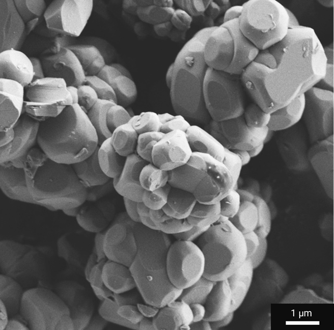
Capture Nanoscale Surface Detail with Confidence
Capture nanoscale surface detail with confidence. BrightBeam™ optics reveal the intricate features of battery materials with exceptional clarity and resolution.
Analyze Beam-Sensitive Materials Safely
Preserve the surface integrity of delicate separators and electrodes using high-resolution, low-voltage imaging and cryo-ready operation.
Visualize Battery Interfaces with High Compositional Sensitivity
Enhance contrast at electrode interfaces and identify binder and conductive additive distribution across complex layers with three-angle or energy-filtered BSE detection.
Correlative Insights in a Single System
Reveal the full story of your battery materials with integrated EDS, EBSD, and Raman spectroscopy for a complete picture of composition, structure, and performance.
Simplify Your Workflow with Custom Automation
Streamline QA/QC and Failure Analysis routines using Essence™ Image Snapper, VisualCoder™ programming, and SEM Expert PI scripting for fast, reproducible, operator-independent results.
Protect Sensitive Samples During Transfer and Analysis
Protect air- and beam-sensitive battery materials with an inert gas transfer workflow and cryogenic conditions, ensuring safe, contamination-free handling and imaging.
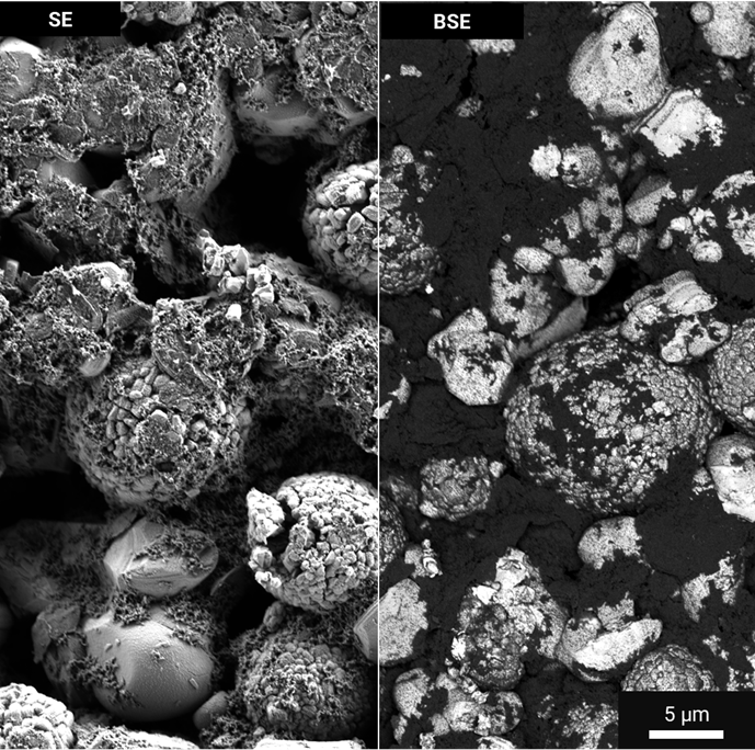
Automated ultra-high-resolution SEM imaging enables fast, quantitative insights into particle size, shape, and surface morphology — all critical to optimizing lithium-ion battery manufacturing.
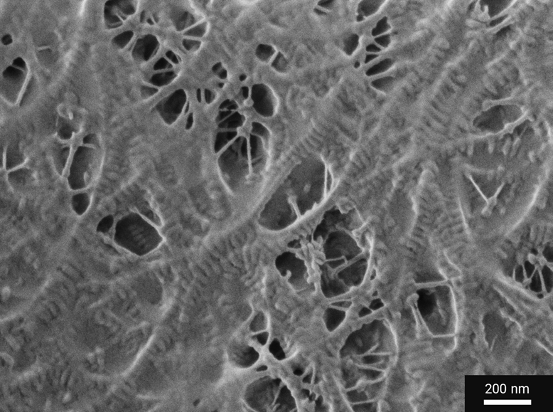
Tescan CLARA delivers non-destructive, nanometer-scale insights into separator porosity — enhancing safety and quality control in lithium-ion battery production.
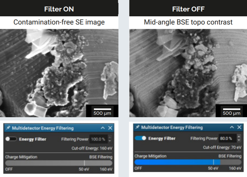
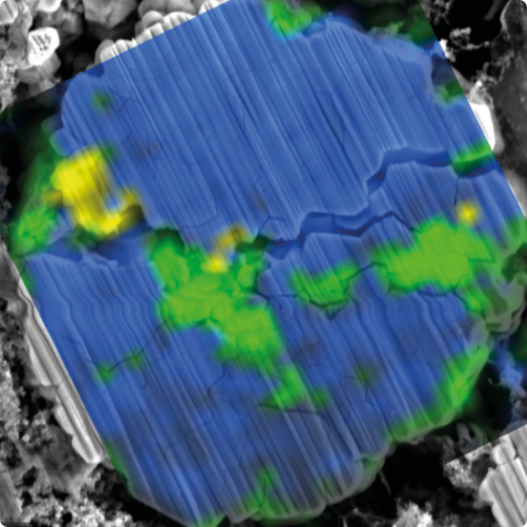
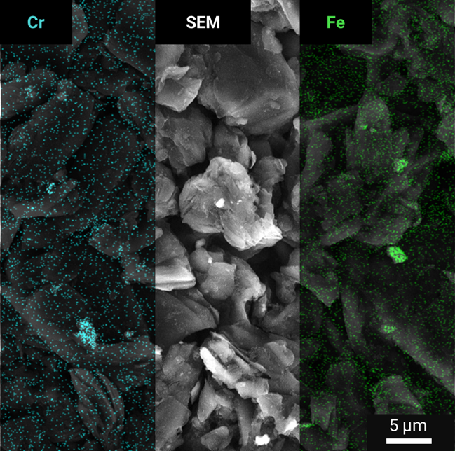
BrightBeam™ technology in Tescan CLARA delivers exceptional beam stability and signal efficiency for ultra-high-resolution imaging at low accelerating voltages. This capability ensures consistent, high-quality imaging conditions for active material analysis, separator evaluation, electrode surface and cross-sectional mapping, interface studies, and SEI characterization across all stages of battery research, quality control, and failure analysis.
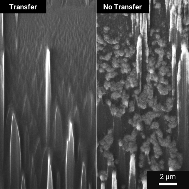
An optimized beam path and detector geometry improve signal-to-noise ratio and surface contrast, even below 1 kV. With its field-free column and In-Flight Beam Tracing™, BrightBeam™ maintains sharp, stable resolution while minimizing charging effects and drift.
This is more than information; it's an advantage. We've compiled our technical whitepapers, detailed product flyers, and on-demand webinars to provide you with the knowledge that makes a real difference. Sign up now to access the insights you need to make an impact.
|
Electron Optics & Imaging |
Electron Source: High-brightness Schottky FEG |
|---|---|
|
Electron Column: BrightBeam™ field-free UHR technology |
|
|
In-Column Detection System: Energy-Filtered Multidetector, Axial |
|
|
High Vacuum Resolution:
|
|
|
Magnification Range: 1× to 2,000,000× |
|
|
Vacuum System |
High Vacuum Mode |
|
Low Vacuum Mode (MultiVac) including N2 or H2O: Up to 500 Pa |
|
|
Stage & Chamber |
5-Axis Motorized Stage (X, Y, Z, rotation, tilt) |
|
Max Sample Size: 335 mm × 280 mm × 133 mm |
|
|
Max Load Capacity: Up to 8 kg |
|
|
Max Load Capacity: Up to 8 kg |
|
|
Chamber view (IR) camera |
|
|
Navigation & Protection |
Wide Field Optics™ (SEM navigation from 1×) |
|
Essence™ 3D Collision Model: Live chamber visualization and collision protection |
|
|
Software & Automation |
Essence™ Software Interface: Customizable with multi-user support |
|
Automated Beam Alignment Routines (e.g., autofocus, beam alignment) |
|
|
Automatic Optimization of Beam Conditions: In-Flight Beam Tracing™ technology |
|
|
VisualCoder™ and SEM Expert PI: Designed for tailored analytical workflows |
|
|
Advanced Analytical Tools |
Energy-Dispersive X-ray Spectroscopy (EDS): Elemental analysis |
|
Electron Backscatter Diffraction (EBSD): Crystal structure and orientation study |
|
|
Raman Spectroscopy: Identifying chemical bonds, crystal structures, stress/strain states, and phase composition |
|
|
Protection of Delicate Samples |
Inert Gas Transfer System for Contamination-Free Sample Handling: Transfer of air- and moisture-sensitive samples between the glove box and external instruments |
|
Cryo-Stage: Utilization of cryogenic conditions to protect sensitive samples during SEM imaging |
|
Electron Optics & Imaging |
|---|
|
Electron Source: High-brightness Schottky FEG |
|
Electron Column: BrightBeam™ field-free UHR technology |
|
In-Column Detection System: Energy-Filtered Multidetector, Axial |
|
High Vacuum Resolution:
|
|
Magnification Range: 1× to 2,000,000× |
|
Vacuum System |
|---|
|
High Vacuum Mode |
|
Low Vacuum Mode (MultiVac) including N2 or H2O: Up to 500 Pa |
|
Stage & Chamber |
|---|
|
5-Axis Motorized Stage (X, Y, Z, rotation, tilt) |
|
Max Sample Size: 335 mm × 280 mm × 133 mm |
|
Max Load Capacity: Up to 8 kg |
|
Max Load Capacity: Up to 8 kg |
|
Chamber view (IR) camera |
|
Navigation & Protection |
|---|
|
Wide Field Optics™ (SEM navigation from 1×) |
|
Essence™ 3D Collision Model: Live chamber visualization and collision protection |
|
Software & Automation |
|---|
|
Essence™ Software Interface: Customizable with multi-user support |
|
Automated Beam Alignment Routines (e.g., autofocus, beam alignment) |
|
Automatic Optimization of Beam Conditions: In-Flight Beam Tracing™ technology |
|
VisualCoder™ and SEM Expert PI: Designed for tailored analytical workflows |
|
Advanced Analytical Tools |
|---|
|
Energy-Dispersive X-ray Spectroscopy (EDS): Elemental analysis |
|
Electron Backscatter Diffraction (EBSD): Crystal structure and orientation study |
|
Raman Spectroscopy: Identifying chemical bonds, crystal structures, stress/strain states, and phase composition |
|
Protection of Delicate Samples |
|---|
|
Inert Gas Transfer System for Contamination-Free Sample Handling: Transfer of air- and moisture-sensitive samples between the glove box and external instruments |
|
Cryo-Stage: Utilization of cryogenic conditions to protect sensitive samples during SEM imaging |
Tescan
Libušina třída 21
623 00 Brno
Czech Republic
No distributors found.