Reveal Sub-Micron Battery Defects with Precision
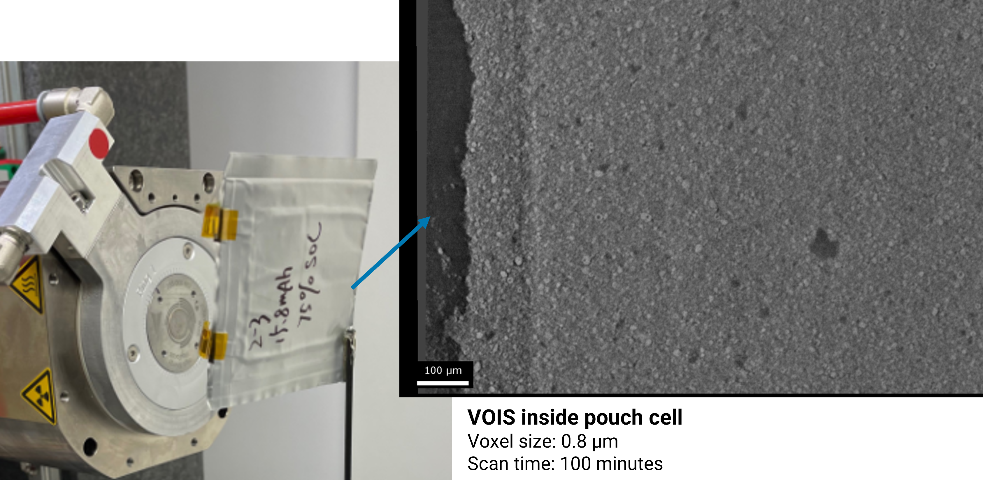
Detect cracks, voids, delamination, and contamination in assembled or cycled cells using sub-600 nm spatial resolution micro-CT. Identify critical failure points before they impact performance.

Advance battery innovation with Tescan UniTOM® HR for Batteries — a sub-micron micro-CT system combining dynamic 4D CT and multiscale imaging in one platform. Purpose-built for in situ testing, defect analysis, and high-throughput QC, it delivers non-destructive insight from full pouch cells to individual electrode particles.

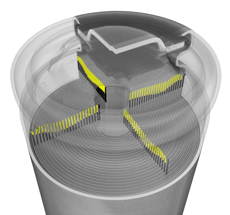
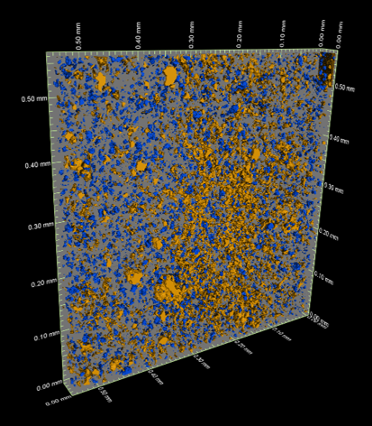
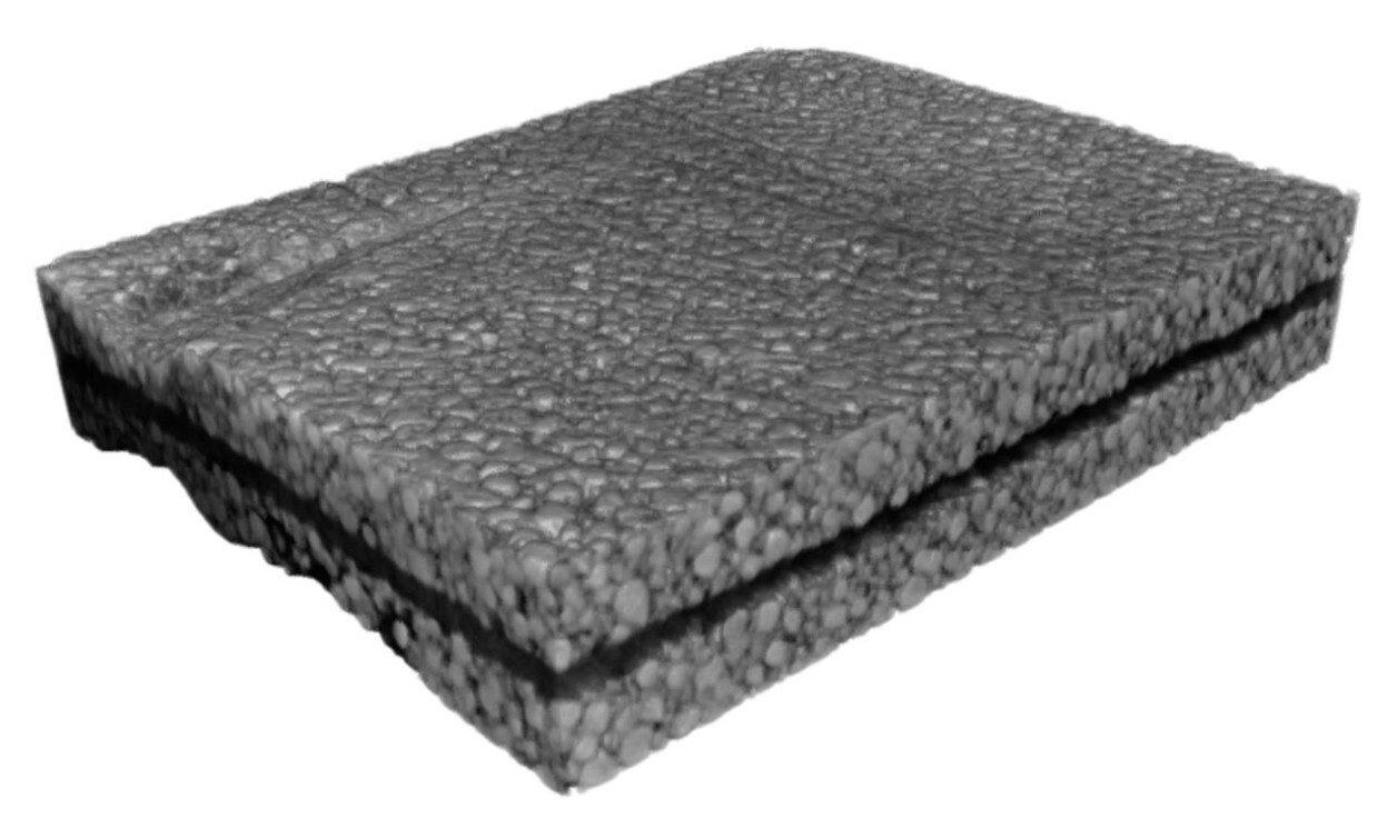
Non-destructive micro-CT imaging uncovers structural voids and packing uniformity — key factors for optimizing battery performance and electrolyte flow.
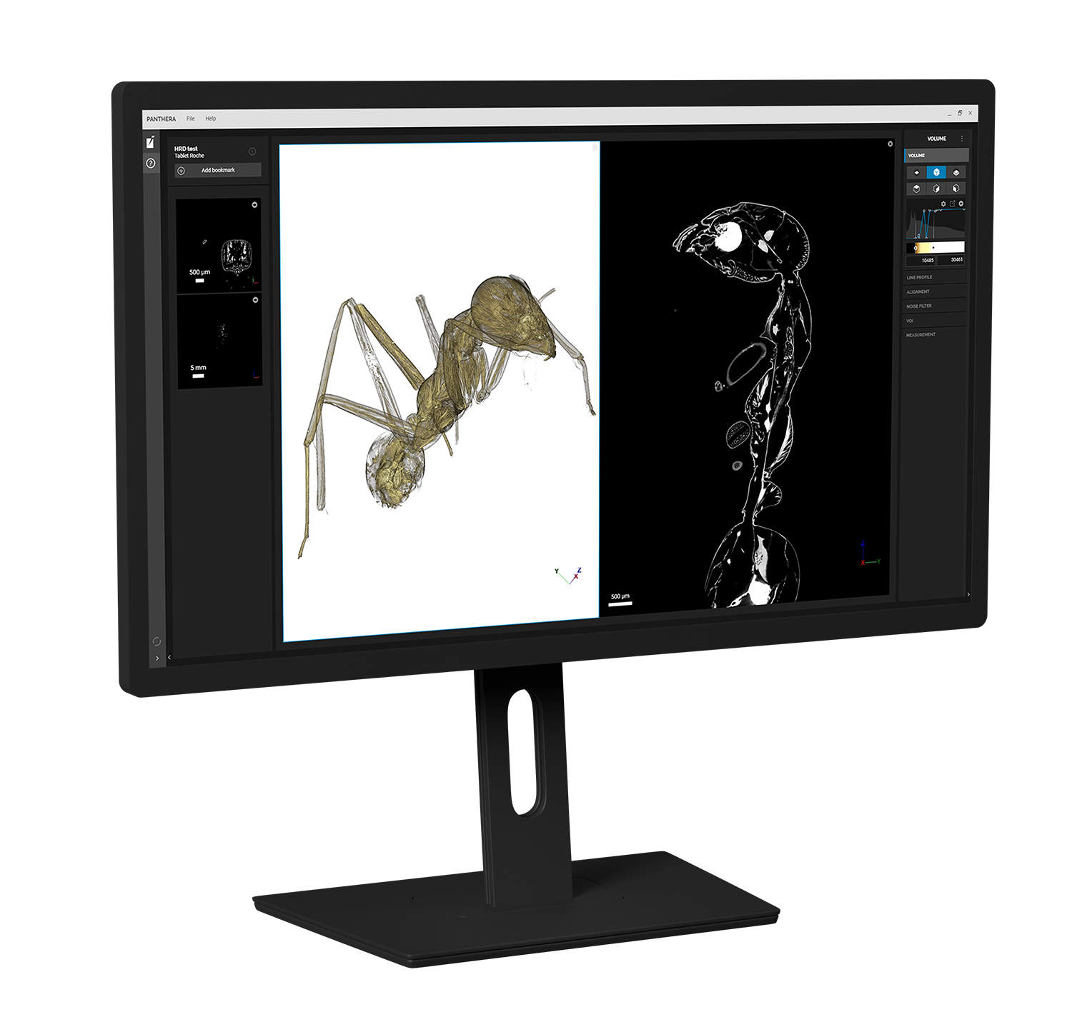
Panthera™ delivers automated 3D and 4D reconstruction with GPU-optimized speed and integrated visualization tools. Users can easily explore large datasets, align time-series volumes, and perform flip-point or differential analysis to detect material changes over time. Intuitive VOIS™ selection connects analysis directly to the scanner for multiscale zoom-in imaging, while advanced filtering tools enhance signal-to-noise ratio and overall image quality.
TEM AutoPrep™ Pro automates the complete TEM lamella preparation process, delivering clean, Ga-free specimens optimized for nanoscale battery analysis. Integrated into the Essence™ GUI, AutoPrep™ Pro removes operator variability and ensures reproducibility across multiple samples and sites.
AI-guided routines handle trench milling, lift-out, grid placement, and 500 eV final polishing, producing lamellae with the right thickness and orientation for high-resolution STEM, EDS, and ToF-SIMS correlation. Multi-site batch operation enables unattended overnight preparation, saving valuable operator time.
With TEM AutoPrep™ Pro, battery researchers and manufacturers can depend on consistent, high-quality lamellae that reduce preparation time and accelerate the investigation of critical interfaces and degradation mechanisms.
.jpg?hsLang=en)

.png?width=1230&height=1293&name=MICRO_UniTOM_HR_1%20(1).png)

-2.png?hsLang=en)