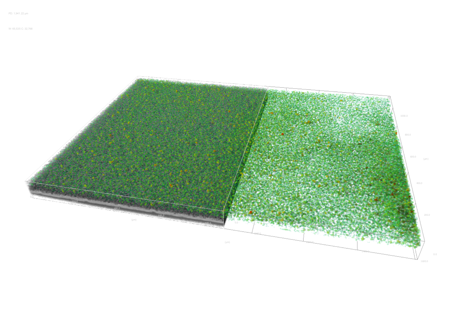
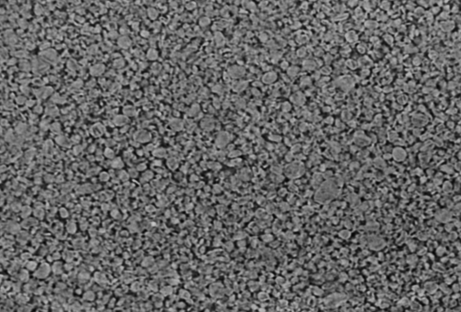
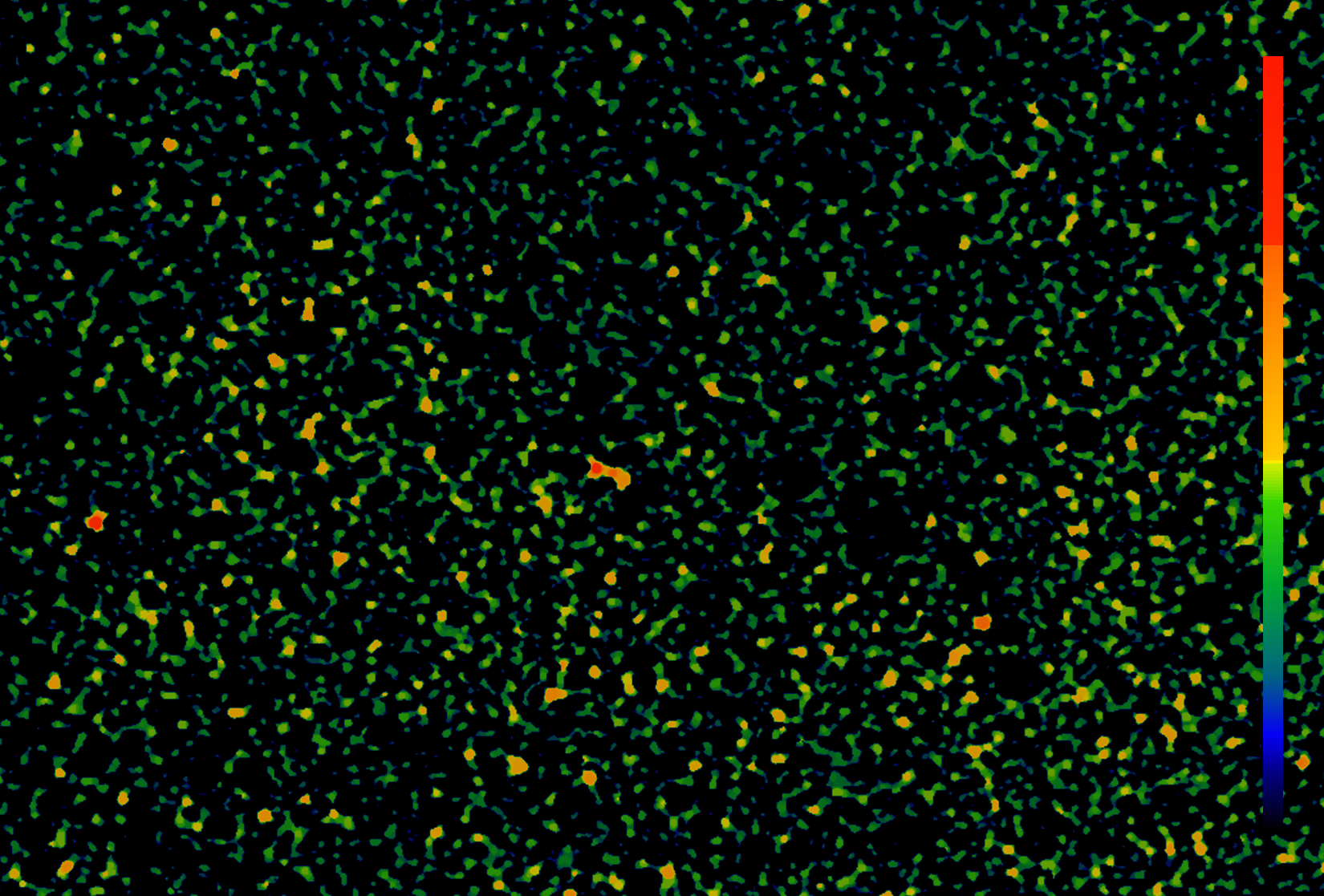
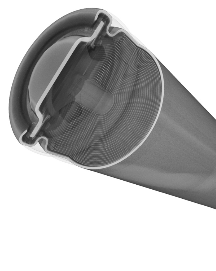
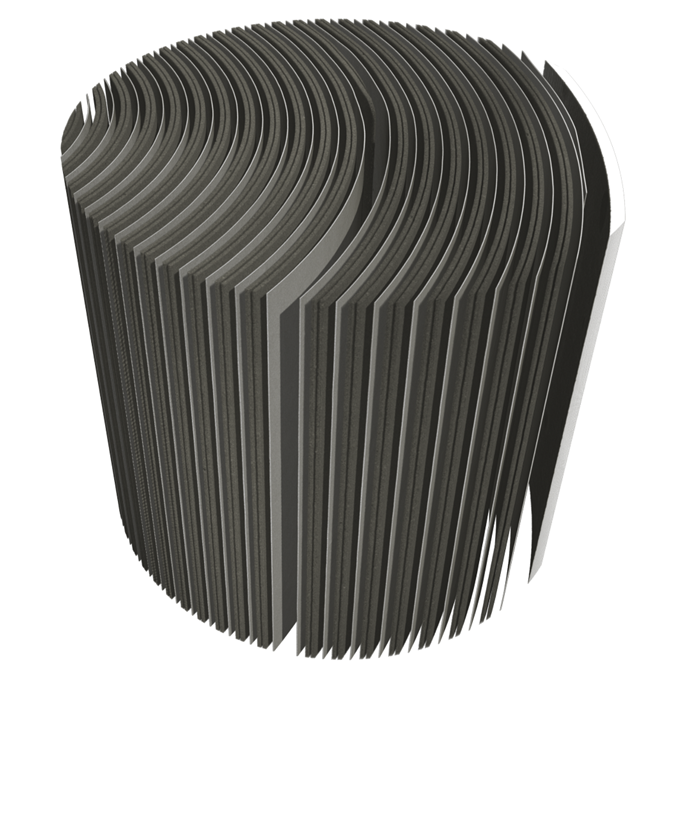
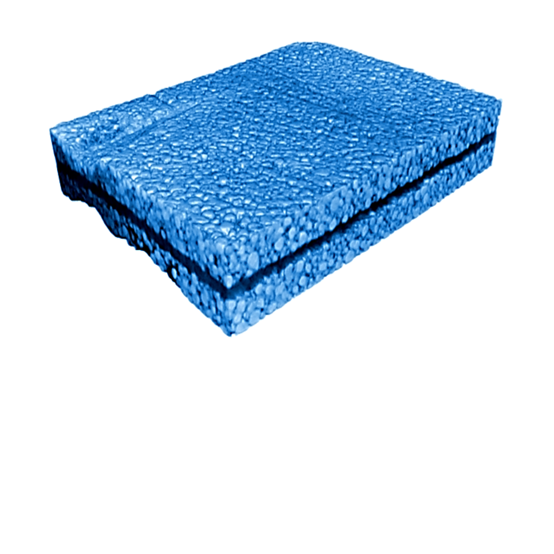
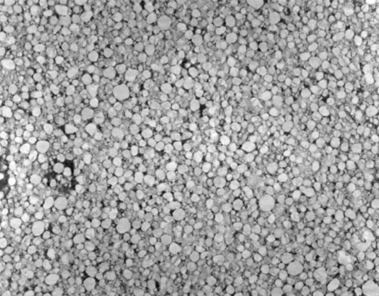
WEBINAR | Up to 40% Faster Automated TEM Lamella Preparation with new Ga⁺ FIB Column

Revealing Electrode Porosity in 3D with Tescan UniTOM HR

Non-destructive micro-CT imaging uncovers structural voids and packing uniformity — key factors for optimizing battery performance and electrolyte flow.

hero
hero
Unlocked content

Porosity in Battery Electrodes: A Hidden Variable with Major Impact

Porosity is more than empty space. It defines how lithium-ion batteries breathe, cycle, and age. This void fraction controls electrolyte infiltration, affects internal resistance, and impacts how well the cell handles fast charging or long cycles.

In this study, Tescan applies UniTOM HR micro-CT to investigate porosity in commercial lithium-ion electrodes. From full battery packs down to individual graphite particles, multi-scale Volume of Interest scanning and high-speed 3D reconstructions offer deep insight — without cutting or unpacking a single cell.

Why Study Electrode Porosity

with Tescan

01
Root of the Problem

Why Porosity Is a Critical Metric in Battery Manufacturing

 Whether designing cells for energy density, fast charging, or cycle stability, porosity plays a defining role. The pores in battery electrodes affect how much electrolyte a cell can hold, how ions move, and how well the electrode bonds to current collectors.

Too much porosity and mechanical strength suffer; too little and ionic resistance rises. Optimizing this structure is central to both performance and safety — yet traditional tools often can’t visualize it in 3D. Enter micro-CT: a powerful, non-destructive method for analyzing porosity at the scale where it matters most.

02
Materials and Methods

MultiScale Micro-CT with Automated VOI Targeting

An industrial lithium-ion battery was first scanned at 15 µm voxel size, covering the entire cell in four vertical sub-scans. Using Tescan Panthera™, these were merged automatically. Volume of Interest (VOI) regions were then selected for higher-resolution re-scanning.

For detailed analysis, a graphite anode foil was isolated and imaged at 1.0 µm voxel size using Tescan UniTOM HR. All reconstructions were performed in Panthera™, with pore segmentation and analysis conducted in Dragonfly 3D World software.

03
Results and Discussion

From Electrode Packing to Pore Size Distribution

 Initial scans of the full cell resolved all major layers — anode, cathode, sleeve, and collector — allowing macro-level defect inspection. At higher resolution, micro-CT revealed clear 3D separation between graphite particles and pores, enabling direct porosity measurements. The observed average porosity was below 30%, with pore diameters ranging from 1.2 to 9.6 µm.

This even distribution is ideal for effective electrolyte infiltration and helps reduce production delays. Incomplete packing and particle delamination were also visible; these are potential indicators of mechanical stress or production errors. This workflow supports both process validation and performance optimization in real time.

GET IN Touch

Contact us

Get the most out of Tescan

This is more than information; it's an advantage. We've compiled our technical whitepapers, detailed product flyers, and on-demand webinars to provide you with the knowledge that makes a real difference. Sign up now to access the insights you need to make an impact.

Tescan Instruments & Technology

Used in This Workflow

Tescan UniTOM HR

Tescan UniTOM HR offers non-destructive, multiscale imaging for battery cells, modules, and materials — from macro inspection to sub-micron porosity analysis.

  • Sub-micron voxel resolution with high-speed scanning
  • Multiscale Volume of Interest (VOI) workflows for targeted analysis
  • Ideal for evaluating electrodes, separators, and full cell assemblies 
MICRO_UniTOM_HR_1-2

GET IN Touch

Contact us

map

Where can you find us:

Tescan Brno
Libušina třída 21
623 00 Brno
Czech Republic

info@Tescan.com