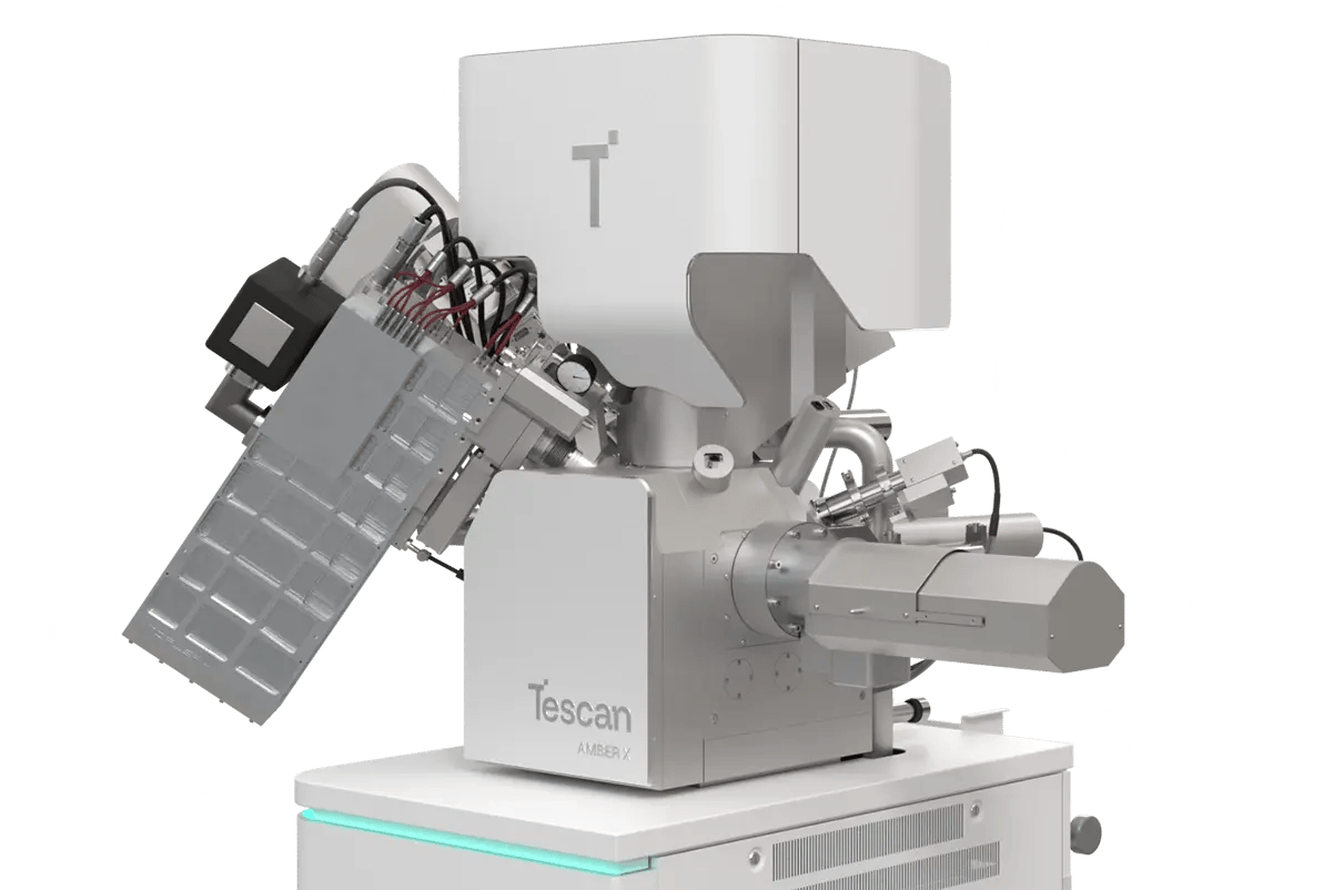
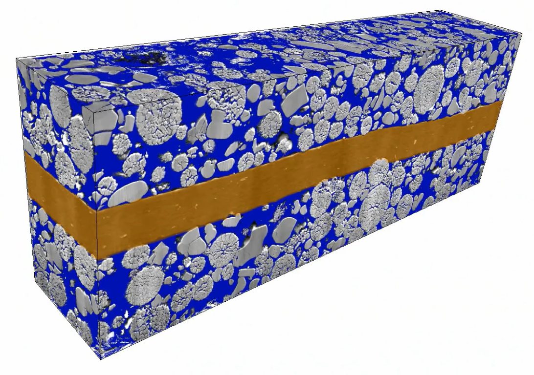
WEBINAR | Up to 40% Faster Automated TEM Lamella Preparation with new Ga⁺ FIB Column

Quantify Battery Electrode Porosity Across Large Volumes with Tescan AMBER X 2

Measure electrode porosity, pore connectivity, and accessibility in 3D using high-volume FIB-SEM tomography.

hero
hero
Unlocked content

Correlate Electrode Structure with Battery Performance, Lifespan, and Manufacturing Quality

Battery electrodes often exhibit heterogeneous porosity with pore networks, and particle detachment, all of which influence electrolyte infiltration and cycling behavior. Surface SEM and 2D cross sections offer only partial views, missing the 3D complexity and structural variation that impact performance and quality.

Tescan AMBER X 2 offers a high-throughput, large-volume FIB-SEM tomography workflow that delivers detailed 3D insights into pore morphology, porosity, and structural uniformity.

Evaluate real electrode behavior across statistically meaningful volumes without relying on assumptions. 

Why Evaluate Battery Electrode Porosity

with Tescan AMBER X 2

01
Root of the Problem

Why Traditional Methods Miss Critical Porosity Behavior in Battery Electrodes

Understanding how electrode porosity affects battery performance requires visibility into internal pores, pore connectivity, and particle detachment. Standard techniques like 2D SEM cross-sectional analysis only reveal partial information. They miss the spatial variation and structural inhomogeneities that drive electrochemical behavior.

Tescan AMBER X 2 enables high-throughput 3D FIB-SEM tomography to evaluate electrode morphology across large volumes with nanoscale resolution and full structural context.

  • Large-volume imaging for statistically relevant data
  • 3D porosity mapping across full electrode cross sections
  • Quantitative porosity and connectivity analysis
  • Correlation of structure with electrolyte infiltration and cycling performance

Characterize battery electrode porosity in true 3D using Tescan AMBER X 2 without missing the structural details that matter most. Enhance your understanding of electrode composition by embedding elemental and chemical information from advanced analytical techniques like EDS and ToF-SIMS into the 3D FIB-SEM tomography workflow. 

02
Materials and Methods

How Electrode Porosity Was Characterized Using Tescan AMBER X 2

Battery electrodes were examined in their original, unsanctioned state to preserve structure and ensure representative analysis. A cross-sectional region of graphite anode was selected for large-volume FIB-SEM tomography using Tescan AMBER X 2.

Automated Xe plasma FIB milling enabled high-throughput serial slicing across volumes up to 80 µm × 80 µm × 88 µm, while high-resolution SEM imaging captured each layer in fine high detail. This combination allowed for full 3D reconstruction of pore networks and particle connectivity.

Data was processed and visualized using Tescan’s 3D Viewer software, while electrode porosity and pore size distribution were evaluated using Dragonfly 3D World.

With accurate, volumetric insight into real electrode morphology, this workflow supports both R&D and quality control needs.

03
Results and Discussion

3D Visualization of Electrode Porosity and Structural Variation

Tescan AMBER X 2 enabled high-resolution 3D imaging of an intact lithium-ion battery electrode. Large-volume 3D FIB-SEM tomography revealed internal pore networks, porosity, and particle detachment in the graphite anode.

Structural variations across layers, including porosity and particle connectivity, were clearly captured at nanoscale resolution. These features directly influence electrolyte infiltration, charge transport, and cycling stability.

Quantitative datasets provided statistically meaningful comparisons across materials and batches, supporting both design optimization and process quality control.

With this level of volumetric insight, you can uncover how electrode structure drives performance across charge cycles. 

GET IN Touch

Contact us

Get the most out of Tescan

This is more than information; it's an advantage. We've compiled our technical whitepapers, detailed product flyers, and on-demand webinars to provide you with the knowledge that makes a real difference. Sign up now to access the insights you need to make an impact.

Tescan Instruments & Technology

Used in This Workflow

Tescan AMBER X 2

Tescan AMBER X 2 gives you access to high-throughput Xe plasma FIB milling and high-resolution SEM imaging. This allows you to reconstruct large volumes of electrode material in 3D with fine detail.

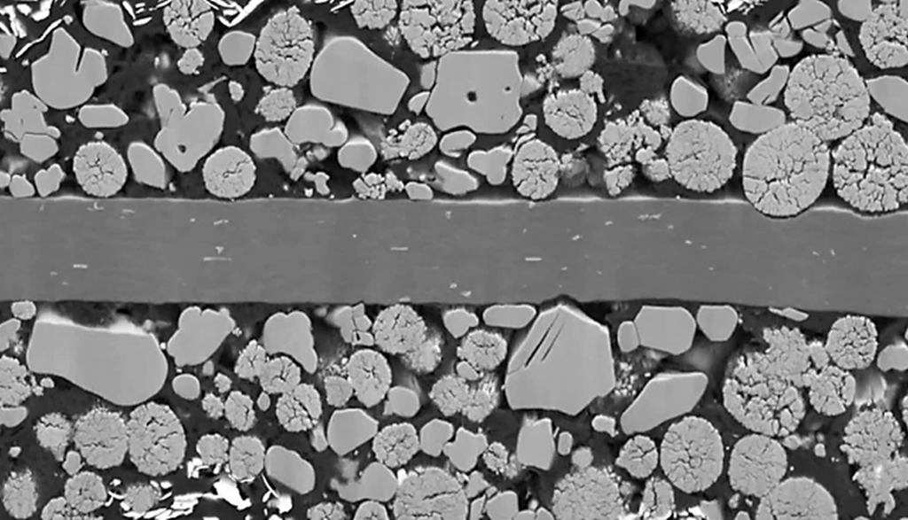
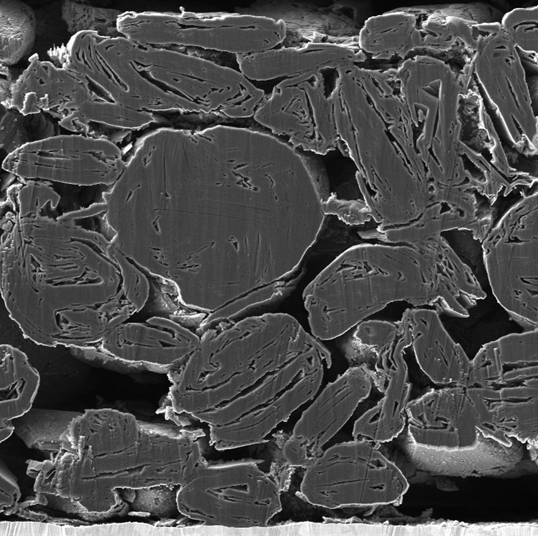
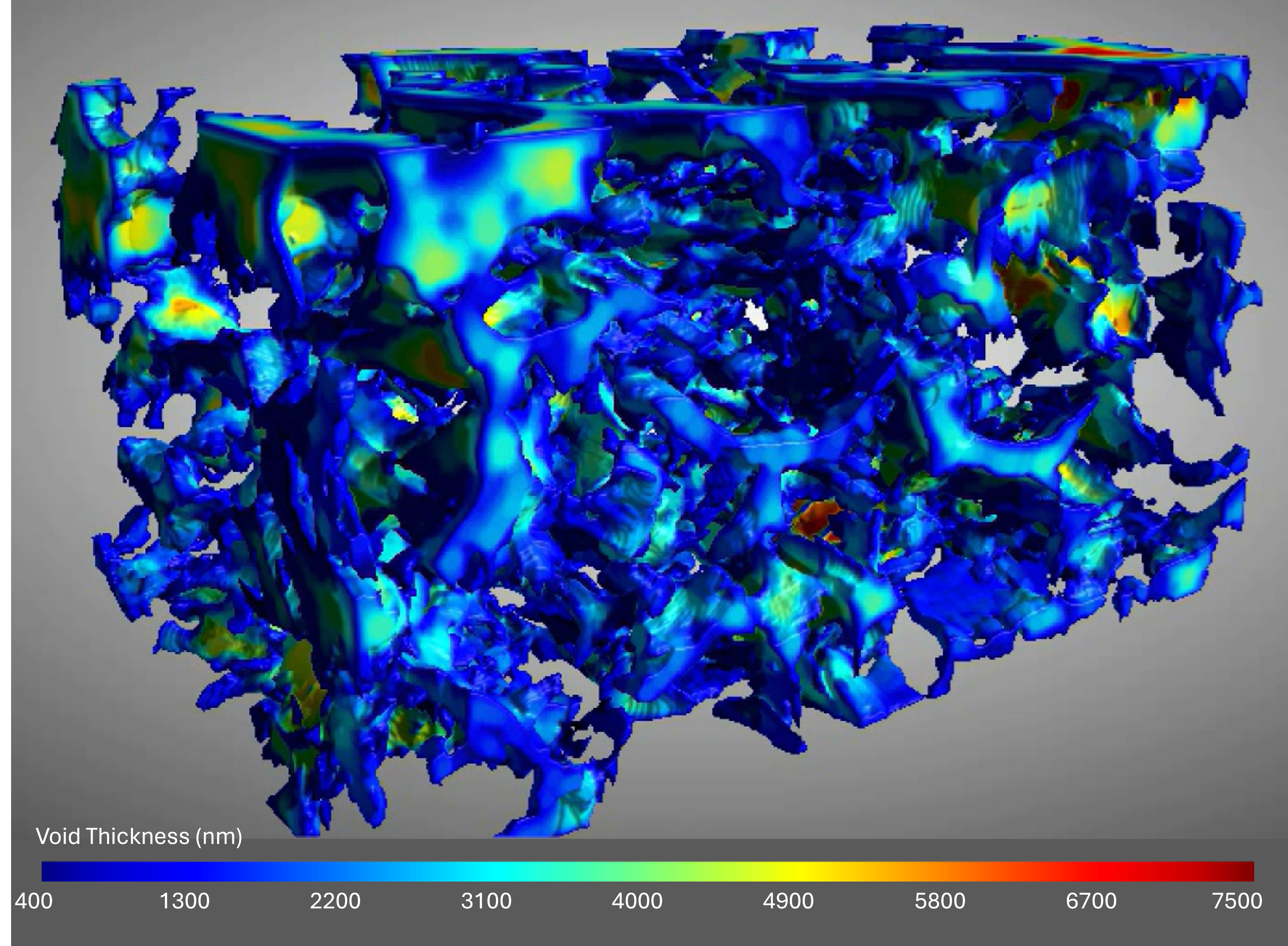
With this workflow, you can visualize pore morphology, porosity, and particle connectivity across entire electrode regions, helping you connect internal structure to performance, quality, and manufacturing outcomes.

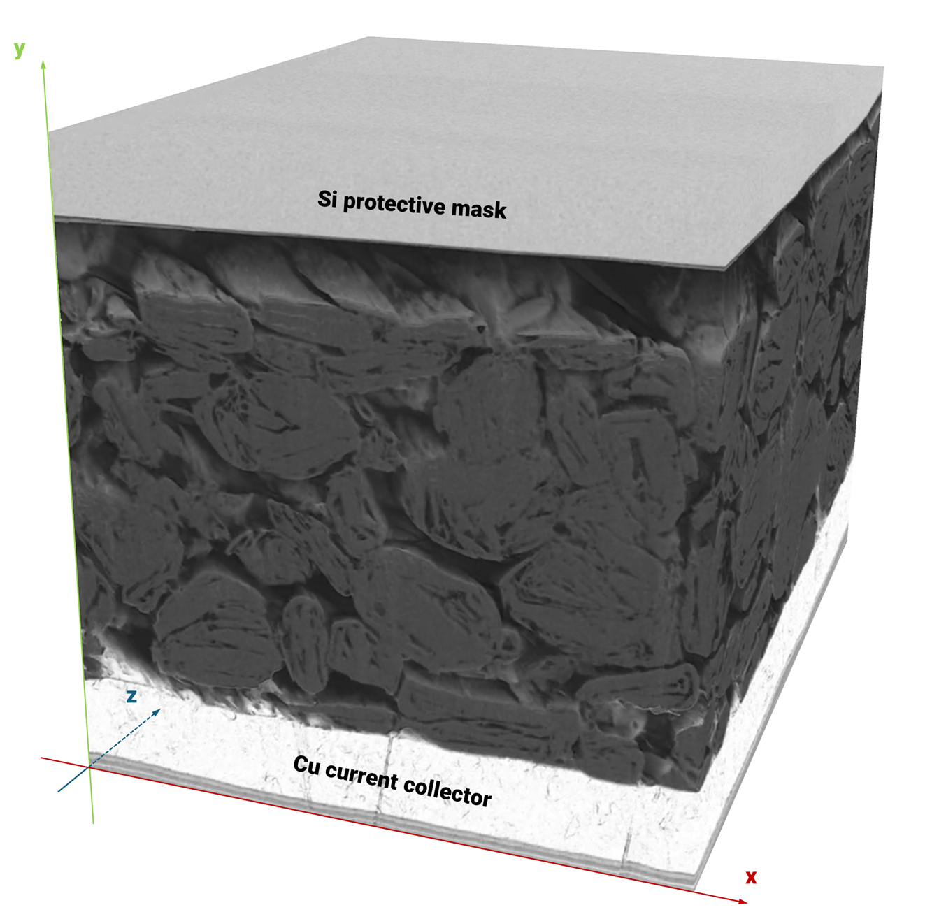
  • High-throughput Xe plasma FIB milling: Enables slicing of volumes up to hundreds of micrometers along all three spatial axes for statistically relevant analysis.

  • High-resolution SEM imaging: Lets you capture nanoscale detail for accurate porosity and structural evaluation.

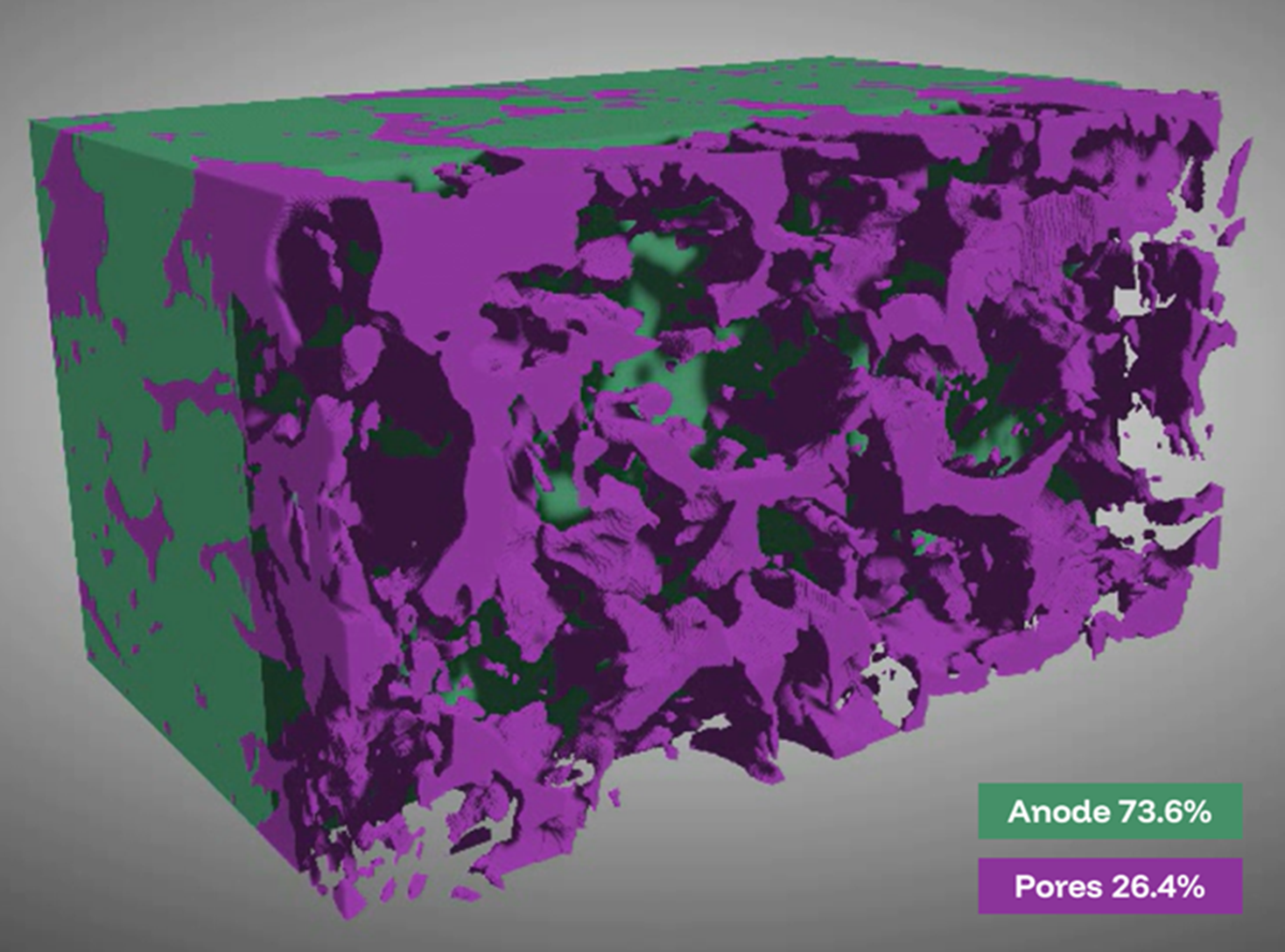
  • Integrated 3D reconstruction: Enables you to generate complete volumetric datasets from slice-and-view data.

  • Multimodal analytical capability: Allows access to multiple complementary analytical techniques, supporting detailed structural and chemical analysis across 3D volumes.
AMBER-X2

GET IN Touch

Contact us

map

Where can you find us:

Tescan Brno
Libušina třída 21
623 00 Brno
Czech Republic

info@Tescan.com