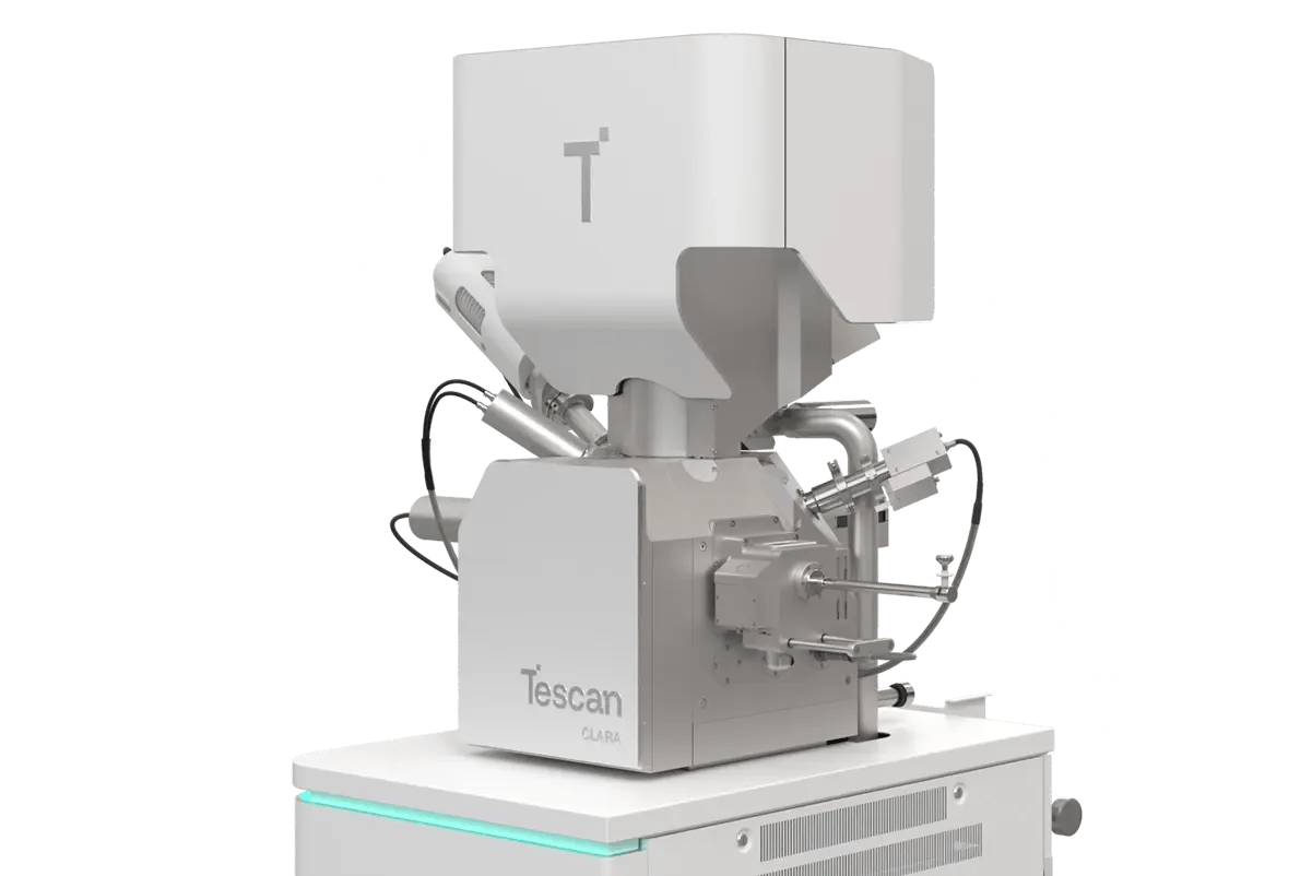
WEBINAR | Up to 40% Faster Automated TEM Lamella Preparation with new Ga⁺ FIB Column

High-Throughput Particle Size Analysis of Battery Powders

Automated SEM imaging with Tescan CLARA reveals morphology and size variation in Ni-based cathode materials. 

hero
hero
Unlocked content

Surface-Level Data That Goes Beyond the Surface

In lithium-ion battery production, not all powder particles are created equal. Variability in particle size and surface texture can influence how well powders flow, how densely they pack, and how uniformly electrodes are coated and calendared.

Using Tescan CLARA ultra-high-resolution SEM in combination with automated imaging workflows, researchers captured detailed surface data across multiple magnifications, enabling both qualitative and quantitative insight into a real Ni-based lithium-ion battery cathode powder.

The entire dataset was acquired in minutes, supporting efficient characterization that can inform quality assessment and materials development.

Why Surface Analysis Matters

for Battery Powders?

01
Root of the Problem

Powder Uniformity Drives Performance

Battery-grade powders must meet demanding criteria not just in their chemistry, but also in size and shape. Inconsistent particle dimensions lead to irregularities in the electrode layer, which can cascade into poor cycle life, reduced capacity, and increased resistance.

Conventional SEM analysis can capture these differences, but it's often too slow, too subjective, or too limited in throughput. A more effective approach combines automated acquisition, nano-resolution imaging, and software-based size extraction, enabling consistent, high-throughput insights that support quality control and materials development. 

02
Materials and Methods

Automated Multiscale Imaging of Cathode Powder

The sample consisted of a Ni-based lithium-ion battery powder, representative of current-generation cathode materials. Three imaging scales were selected: 

01
250 µm field of view for overall distribution
02
50 µm to assess mid-sized particle clusters
03
10 µm for detailed surface texture

Imaging was performed using a Tescan CLARA UHR SEM operated at 1 keV and low beam current. Using Essence™ Image Snapper, the system automatically acquired and optimized each image based on focus, contrast, and scale without operator input.

For size and morphology analysis, image stacks were processed using MIPAR, which extracted diameter distributions and particle shape metrics from binarized image data

03
Results and Discussion

Structural Domains and Phase Interfaces

Analysis revealed particle diameters ranging from 1.2 µm to 10.9 µm, with an average of 4.2 µm ± 1.2 µm. This degree of variability may influence flow characteristics during slurry mixing and affect how the electrode responds to calendaring.

High-magnification images showed clusters of smaller primary particles fused at grain boundaries, creating increased surface area, but also potential hotspots for degradation. Because the data was acquired at multiple magnifications, users could correlate global size trends with local texture effects.

This approach demonstrates how SEM-based surface analysis can deliver not just snapshots, but also insights into the links between particle design and electrochemical performance.

GET IN Touch

Contact us

Get the most out of Tescan

This is more than information; it's an advantage. We've compiled our technical whitepapers, detailed product flyers, and on-demand webinars to provide you with the knowledge that makes a real difference. Sign up now to access the insights you need to make an impact.

Tescan Instruments & Technology

Used in This Workflow

Tescan CLARA

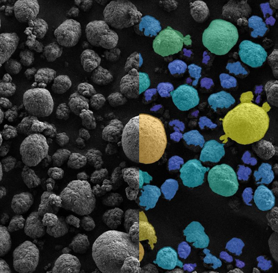
A field emission scanning electron microscope optimized for low-kV imaging and high-contrast surface visualization.

  • Delivers nanometer resolution at low accelerating voltages

  • Ideal for beam-sensitive and nano-structured materials

  • Consistent performance across large samples and multiple scales

CLARA GM Mat. Science

Tescan Essence™ Image Snapper – Automated SEM Imaging Workflow

Project-based automation software that manages image acquisition across multiple fields of view.

  • Unattended SEM imaging with automated contrast and focus adjustment

  • Reduces operator variability and improves reproducibility

  • Tailored for QA workflows, materials screening, and high-throughput morphology capture

GET IN Touch

Contact us

map

Where can you find us:

Tescan Brno
Libušina třída 21
623 00 Brno
Czech Republic

info@Tescan.com