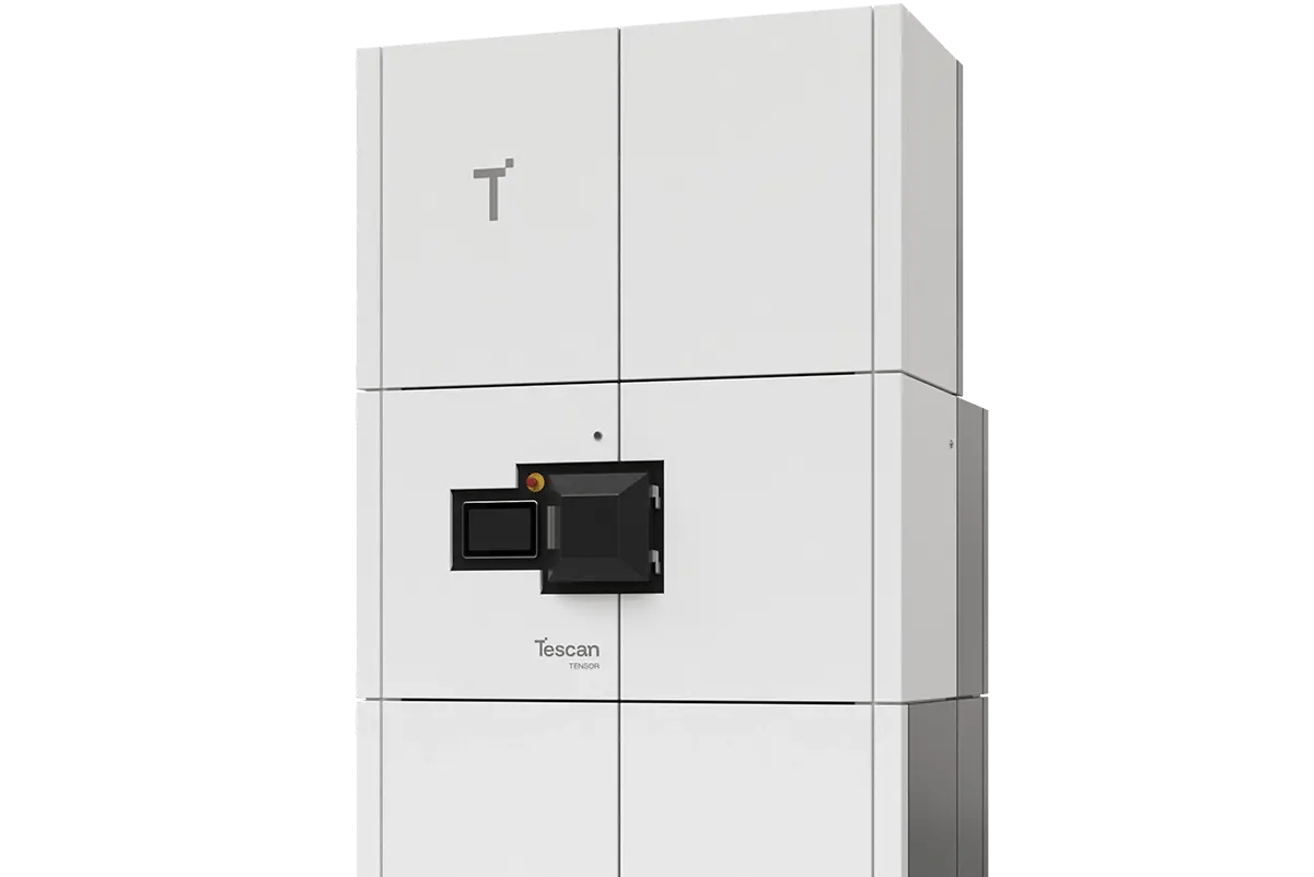
WEBINAR | Dynamic-to-Detail Micro-CT in One Workflow: UniTOM HR 2

Atomic Structure Determination Using Precession Electron Diffraction Tomography (PEDT)

Solve crystal structures including hydrogens at sub-Angstrom resolution with precession-assisted 3D-ED workflow on Tescan TENSOR.

hero
hero
Unlocked content
Precession electron diffraction tomography (PEDT) with Tescan TENSOR enables accurate 3D structural analysis of sub-micron crystals. By combining stepwise 3D ED data collection with beam precession and optional 4D-STEM mapping at each tilt step (5D STEM), it delivers high-quality diffraction data suitable for both kinematical and dynamical refinement — even for challenging beam-sensitive samples.

Using Precession Electron Diffraction Tomography (PEDT)

for Atomic Structure Determination

01
Root of the Problem

Structure Determination from Nanocrystals Remains Challenging

Nanocrystalline materials are commonly used in geology, pharmaceuticals, and materials science. However, solving their atomic structures remains challenging. While traditional X-ray diffraction (XRD) techniques require crystals few microns in size, especially when embedded in complex matrices, TEM-based electron diffraction techniques (micro-ED) are affected by strong dynamical scattering. This distorts diffraction intensities and limits the accuracy of structural refinement — particularly in beam-sensitive samples. A successful approach must not only reduce the dynamical scattering effects to maximize data quality, but also track the sample reliably during tilt series acquisition — especially for nanocrystals < 500 nm.

02
Materials and Methods

3D ED Workflow with Tescan TENSOR

Tescan TENSOR employs precession diffraction tomography (PEDT) for structure determination by 3D ED. This benchmark study used natrolite sample that was prepared by dispersing fine powder in isopropanol and drop-casting onto a holey carbon grid. After plasma cleaning, a single ~2 µm crystal was selected for analysis.

Using Tescan TENSOR, electron diffraction tilt series were acquired through stepwise 4D-STEM mapping combined with electron beam precession at a semiangle of 0.5°. The sample was tilted in 1° increments, and at each position a 19 × 16 tile map was recorded.

Only high-quality diffraction tiles were selected and averaged at each tilt step. Tescan’s XYZ reference tracker ensured beam centering and minimized exposure. The hybrid-pixel detector captured patterns at fast readout speed with low background and high contrast.

This approach minimized background from the support film, and distributed the electron dose evenly across the sample — preserving sensitive regions while collecting sufficient data for analysis.

03
Results and Discussion

High-Quality 3D Diffraction Dataset for Reliable Structural Refinement

The acquired dataset was processed using PETS2 software. Lattice parameters and crystal symmetry were assessed, followed by dynamical refinement for atomic structure determination.

The final data showed:

  • Atomic resolution at 0.63 Å
  • 73% completeness from a single dataset across the tilt range
  • Refined R-factor below 8%

These results enabled confident localization and refinement of all atoms, including hydrogens. Constructed difference maps also revealed density of hydrogen atoms, which cannot be obtained by XRD.

This study demonstrates that PEDT with Tescan TENSOR is highly effective for atomic structure determination and analysis of nanocrystals, even when crystals are small, beam-sensitive, or part of complex mixtures.

GET IN Touch

Contact us

Get the most out of Tescan

This is more than information; it's an advantage. We've compiled our technical whitepapers, detailed product flyers, and on-demand webinars to provide you with the knowledge that makes a real difference. Sign up now to access the insights you need to make an impact.

Tescan Instruments & Technology

Used in This Workflow

Tescan TENSOR with integrated DECTRIS QUADRO

Tescan TENSOR is a dedicated analytical STEM platform optimized for 4D-STEM and 3D ED workflows enhanced by beam precession. It features full integration of the DECTRIS QUADRO hybrid-pixel detector to deliver rapid, high-precision diffraction data for materials research.

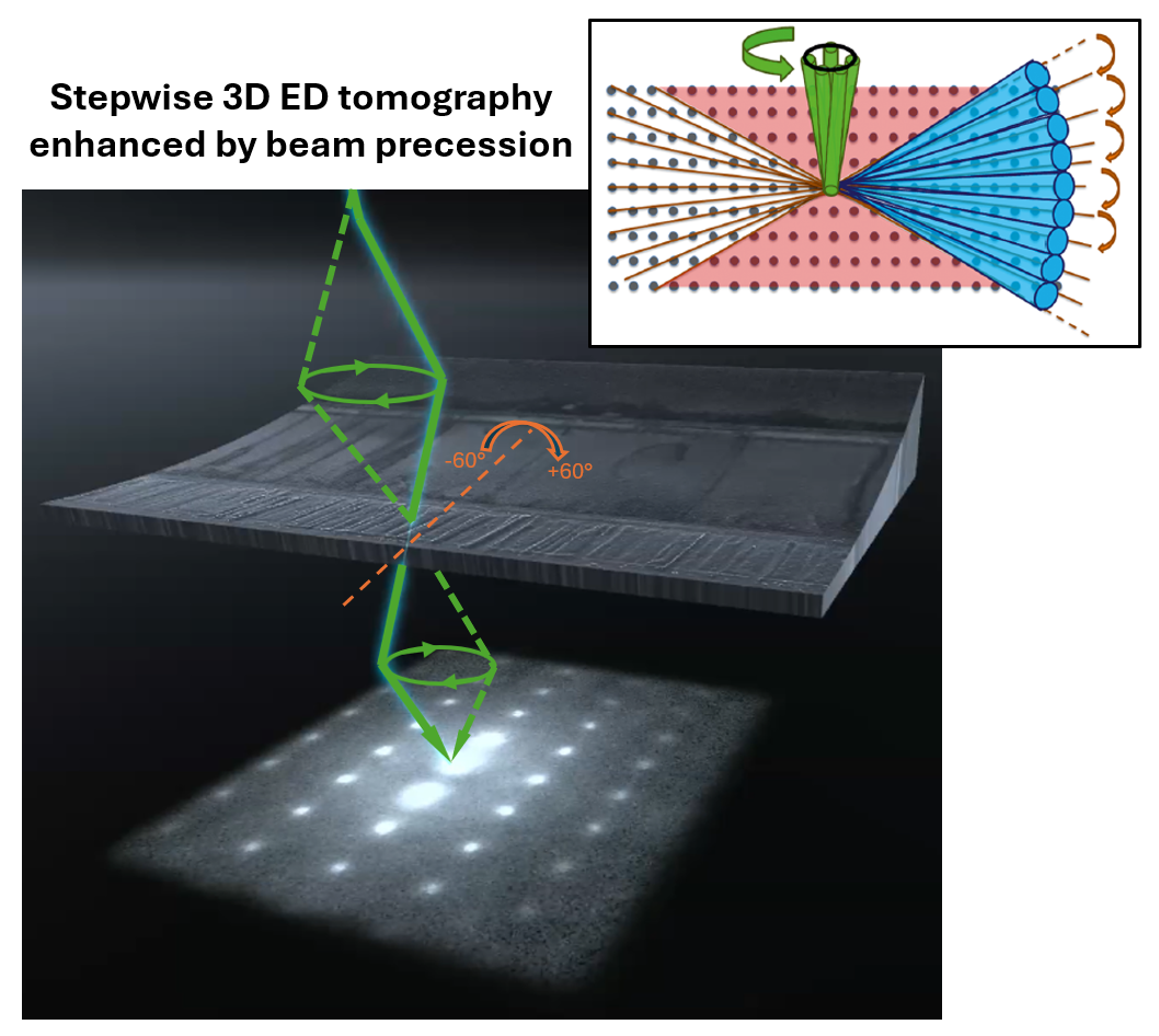
  • Fully integrated beam precession running at 72,000 Hz.

  • DECTRIS QUADRO detector with high dynamic range, single-electron sensitivity, and fast readout speed (up to 4,500 fps)

  • Beam precession for improved data quality due to reduced dynamical effects

  • Streamlined and automated data acquisition with Explore™ user interface and 3D ED data processing with customized PETS software
TENSOR_1

GET IN Touch

with specialist

map

Where can you find us: 

Tescan
Libušina třída 21
623 00 Brno
Czech Republic

130405923 us US 37.09024 -95.712891 25.3575 29.349345 20.67957527 42.082797 39.91384763 -33.693421 13.93320106 3.039986586 31.997988 38.050985 47.579533 48.1485965 58.375799 54.663142 19.195447 56.975106 50.493053 45.868592 10.79556993 44.35660598 43.2371604 55.536415 14.557577179752773 32.100937 -6.116829 -6.212299277967318 23.7104 -33.471062 31.998740087 -23.69149395 43.462349 51.529848 49.1893523 49.197486 25.072375 31.075811 1.299027 40.676979 52.30150662 51.013813 35.684121 37.479653 52.246622 40.581349 39.911632 -26.1811371 41.818215 33.429928 -12.08688

No distributors found.