Intuitive multimodal characterization of samples using the conventional BF/DF STEM imaging, EDS compositional analysis, and 3D STEM/EDS tomography, as well as advanced electron diffraction workflows for 4D-STEM and 3D ED measurements.
Revolutionizing Analytical STEM and 4D-STEM Characterization
Tescan TENSOR™ analytical STEM diffraction microscope offers a new way of TEM/STEM characterization of samples at much higher productivity compared to the traditional TEM/STEM systems on the market. Fast, multimodal characterization of nanoscale morphological, chemical, and structural properties of functional materials, thin films, synthetic particles, and other samples is achieved flawlessly due to due to high levels of automation, either by using the conventional techniques of STEM imaging and EDS compositional analysis or the advanced 4D-STEM and 3D-ED workflows, while diffraction data quality is enhanced by fully integrated and synchronized beam precession.
TEM_%26_4D-STEM_Characterization.webp?width=800&height=700&name=01-Analytical_(S)TEM_%26_4D-STEM_Characterization.webp)
TEM_%26_4D-STEM_Characterization-mobile.webp?width=500&height=500&name=01-Analytical_(S)TEM_%26_4D-STEM_Characterization-mobile.webp)
Multimodal STEM characterization of nanoparticles in carbon nanotubes
Precise identification of both structure and chemistry on the nanoscale can be leveraged to advance research into the next generation of engineering materials, across industry and academia. This Tescan TENSOR™ application note provides an example of multimodal chemical and crystallographic characterization of metal particles encapsulated within carbon nanotubes. By combining STEM imaging, nanobeam diffraction with crystal orientation analysis (4D-STEM) and EDX mapping, we can work towards a comprehensive understanding of this material system. This application note was produced in collaboration with Prof. Andy Brown and Dr. Zabeada Aslam of Leeds University (UK).

Nanoscale Phase Analysis in Battery Electrodes
The differentiation of lithium phosphates in battery anodes is a challenging task due to the relatively similar elemental compositions of the different phosphate phases. In this application note we show how Tescan TENSOR™’s 4D-STEM phase analysis, using precession electron diffraction patterns, helps to improve the differentiation of these phases and identify the penetration of crystalline TiO2 nanoparticles along LiTi(PO4)3 spindle particles. The distribution and relative orientation relationship between different phases in the battery anode can be easily deduced using this analysis. We also show how titanium oxide particles are preferentially distributed along phosphate particle grain boundaries.
.webp?width=1000&height=708&name=Nov%C3%BD%20projekt%20(17).webp)
Enhancing 4D-STEM phase analysis by EDX
TENSOR™ provides natively correlated complementary analytical data due to perfect synchronization and full integration of all hardware modules. In the case of the 4D-STEM datasets, each diffraction pattern can be acquired together with an EDS spectrum, fast and perfectly synchronized. Together, diffraction and spectroscopy data encapsulate the full picture of electron–specimen interaction, from which a wide range of material properties can be derived and some inherent challenges of individual techniques overcome. This technical note shows how EDS signals can improve phase mapping when different phases have almost identical lattice parameters but different composition.
%20(1).webp?width=600&height=439&name=Figure%201(2)%20(1).webp)
Interactive phase and orientation mapping by precession-assisted 4D-STEM
Tescan TENSOR™ provides 4D-STEM capabilities enabling fast nanoscale phase and orientation analysis. In this technical note, the methodology of phase and orientation analysis is described, subjected to drop casted BGO (Bi12GeO20) on evaporated aluminium. Phase and orientation of BGO is measured from a scanning diffraction dataset, by matching the acquired electron diffraction patterns with BGO and Al templates, for each pixel in the dataset. It is shown how precession electron diffraction (PED) improves the confidence of indexing, by enhancing the number of reflections in the diffraction pattern, leading to greater differentiation of the information contained in the patterns acquired from the two phases.
.webp?width=1000&height=580&name=Figure%201%20(2).webp)
Structural characterization of the deformation behavior in alloys
This application note presents a study into Vickers indentation induced plastic deformation in Nickel superalloys. Precession assisted 4D-STEM with automated crystal orientation analysis was used to investigate deformation induced, nanoscale grain re-orientation, in a site-specific FIB prepared specimen. Fast acquisitions, and on-the fly-processing enabled measurements to be optimized, and specimen areas to be localized, in a near real-time iterative manner, which vastly increases measurement and microscope time efficiency.

Enhancing Nanoscale Characterization of Precipitates in Aluminum Alloys by 4D-STEM Phase-Orientation Analysis
This application note demonstrates how the Tescan TENSOR™ analytical STEM facilitates deeper insights into the relationships between aluminum alloy properties and internal structural and compositional features at the nanoscale by multimodal analytical STEM characterization. By combining STEM imaging, EDX compositional mapping, and 4D-STEM phase/orientation analysis, the multimodal analytical results revealed the spatial distribution and internal crystalline structure of strengthening precipitates in the AA2099 alloy, which can be directly linked to structure–property relationships for material and its preparation process optimization.

Structure determination using Precession Electron Diffraction Tomography
Precession electron diffraction tomography (PEDT) with Tescan TENSOR™ enables accurate 3D structural analysis of sub-micron crystals. By combining stepwise 3D ED data collection with beam precession and optional 4D-STEM mapping at each tilt step (5D STEM), it delivers high-quality diffraction data suitable for both kinematical and dynamical refinement — even for challenging beam-sensitive samples.

Structure determination from grains in polycrystalline samples
Tescan TENSOR™ enables material’s structure determination polycrystalline samples by using the precession electron diffraction tomography (PEDT) implementation of 3D ED. Detailed analysis of micro- and nano-scale domains in heterogeneous materials can now be therefore done from a single lamella without the need for preparation of special sample geometries or growing single crystals. The study done in partnership with the group of Prof. Phillipe Boulay from the University of Caen demonstrates how the combination of precession diffraction tomography (PEDT) and electron diffraction mapping (4D-STEM) facilitates the determination of the distribution of stannite and enargite phases in Cu-rich ceramics.
.png?width=2000&name=Figure%202(3).png)
Enabling customized 4D-STEM workflows with open API
Advanced users have the option of open API environment (ExpertPI) for the development of new and custom data acquisition and processing workflows, such as those implemented in the user-friendly streamlined measurement workflows in Explore. Other open-source software packages, such as LiberTEM, py4Dstem or HyperSpy, can be also used in the custom scripts and workflows.
%20(3).webp?width=1000&height=580&name=Figure%202(4)%20(3).webp)
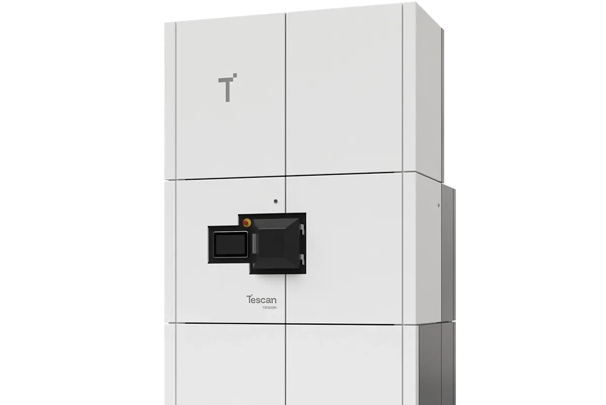
Tescan TENSOR™
A fully integrated analytical scanning transmission electron microscope that captures imaging, diffraction, and EDS data simultaneously for multimodal nanoscale characterization.
- Precession-assisted diffraction for cleaner patterns and better phase indexing
- Real-time orientation and phase mapping with nanometer spatial resolution
- Compatible with large fields of view and automated workflows for battery R&D
Built around your needs
