
Progress in electron microscopy depends on open technical knowledge and experience exchange. That’s why at Tescan, we invite leading researchers to our Brno headquarters to exchange ideas, challenge assumptions, and refine the analytical tools and application workflows that support their work.
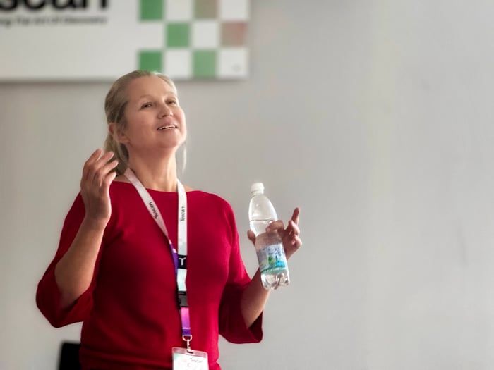
Recently, we welcomed Dr. Tatiana Gorelik, a pioneer in electron crystallography, for a focused session on 3D electron diffraction (3D-ED). Her visit was part of our ongoing commitment to working side-by-side with the scientific community to improve instrumentation and support advanced analytical techniques.
Dr. Tatiana Gorelik’s Perspective on 3D-ED
Dr. Gorelik has more than two decades of experience in TEM and 3D-ED. In her scientific career, she contributed to the development of Automated Diffraction Tomography (ADT) and the processing of electron diffraction datasets for 3D structure determination. Her work supported the evolution of these methods into PETS2, which is widely recognized in academia as a leading software tool for 3D electron diffraction data processing. She was one of the first scientists to determine the structures of organic compounds using 3D-ED. She has contributed to a wide range of scientific fields, including determination of small-molecule crystal structures, 2D electron crystallography, and Pair Distribution Function analysis from electron diffraction data.
Today, she continues her research at the Ernst Ruska-Centre (ER-C) in Germany and remains active in research, educational, and editorial work, serving as an editor for the peer-reviewed journals Acta Crystallographica Section A and Zeitschrift für Kristallographie. To support education in electron crystallography, she is organizing a winter school on electron crystallography in Jülich in March 2026.
Recent Research: from diffraction to imaging through iterative phase retrieval methods
In a recent study, she and Tatiana Latychevskaia (PSI) demonstrated the possibility of direct structure projection reconstruction from a single electron diffraction pattern of MOF nanocrystals. Careful acquisition of the diffraction data allowed for the observation of both low-resolution data, representing the scattering of the nanocrystal shape, and high-resolution data, representing the crystal structure contribution. Both regions could be successfully used to recover direct space information, including the crystal morphology and the projected crystal structure.
Data Collection Strategies for Reliable 3D-ED
During her presentation, Dr. Tatiana Gorelik showed how electron diffraction can solve structures from nanocrystals. She discussed approaches for acquiring diffraction tilt series, such as stepwise acquisition with beam procession and continuous rotation. She described the advantages and disadvantages of each method concerning crystal tracking and the integration of recorded diffraction spot intensities.
She further elaborated on how serial ED datasets and machine-learning models can be used to efficiently determine the unit cell and the atomic structure of highly beam-sensitive samples. She used examples such as organic pigments and pharmaceutical samples to emphasize that the success of a structure solution depends ultimately on data acquisition strategy and associated data quality.
TENSOR Evaluation: Throughput and Small-Crystal Performance
TENSOR was discussed as a platform with clear potential for more automated and higher-throughput data acquisition, especially when combining small 3D-ED tilt series with 4D-STEM data acquisition. Dr. Gorelik noted that this approach can facilitate the efficient collection of diffraction data from a large number of small crystals. She also noted the higher quality of data from very small (<300 nm) crystals compared to traditional electron diffractometers and TEM/STEM microscopes. Ultimately, this helps researchers to capture reliable diffraction data even from challenging samples.
Practical Test: Structure Determination of the Orange Pigment
During her visit, Dr. Gorelik tested TENSOR on an orange pigment sample known for producing tiny, highly agglomerated crystals. Its exact 3D structure has therefore remained unknown for several decades. She collected multiple small 3D-ED tilt-series from individual small crystals, using the small parallel beam size generated by TENSOR, extracted the unit-cell parameters, and used them to index serial 4DSTEM high-signal diffraction patterns.
This combined dataset provided sufficient crystallographic information to solve the atomic structure, which had long been elusive using traditional structural methods. Her measurements showed that TENSOR can efficiently probe crystals at this scale by enabling very small (<100 nm) parallel illumination probe beams, accurately aligned and fast beam precession, and flexible acquisition of diffraction data.
In Conversation: Dr. Tatiana Gorelik on the Future of 3D-ED
We spoke with Dr. Gorelik about her scientific path, her current research, and where she sees the field heading.
What drew you to electron crystallography, and how has the field evolved since you began?
Dr. Gorelik described how her path into electron crystallography began with chemistry, photography, and a moment of chance: “I studied chemistry. At the time, I was doing a lot of photography as a hobby. The combination of image-making and molecular structure guided me toward a career in electron microscopy. I was walking through all the labs that were offering positions for master's students for diploma work. And then there was this lab with a photo laboratory at that time, and it was an electron microscopy lab. So that was a clear choice, and I completed my diploma in electron microscopy.”
She added: “I studied chemistry with a strong focus on organic chemistry, and later moved toward physics and electron microscopy. After years in semiconductor physics, I joined a chemical institute in Mainz, where my original background could come to light, chemistry, together with electron microscopy. So that's how it started. That's why the first materials we looked at were organic molecules."
Her connection to the field is personal: "I feel very much at home with organic materials, with electron crystallography, because it's exactly my vision of the world, of nature. I want to see the atoms, but the atoms have to be arranged in a proper way. Electron crystallography provides the means for me to see what I want to see."
Reflecting on how the field has developed, she recalls her early work in Mainz with Ute Kolb and mentions Ingrid Voigt-Martin and Doug Dorset. She noted, "I was following the complete path of the development and rise of electron diffraction tomography techniques. At that time, electron crystallography was a branch of electron microscopy, and everything in microscopy is related to the zone axis. The approach was to record the zone axis patterns of these organic crystals in different orientations and then try to combine them. It was very aesthetic but extremely tedious to tilt, especially organic crystals, which decay under the beam. So, it required a lot of training."
Progress came with automation: "We then developed automated rotation. Instead of relying on zone-axis patterns, researchers could collect data at constant tilt increments. This shift began in 2005–2006 in Mainz, and our first paper came out in 2007. Many groups have started working on it now, and it's really great to see how it's evolving."
What are the biggest challenges in 3D-ED today, and how can instrumentation help overcome them?
"There are technical challenges. And there are infrastructural challenges. Technical issues include small or disordered crystals, managing large datasets, uncertainty in data processing, and aligning 3D data. They can be tackled either by companies or by us scientists. So, this is one group of problems."
The second challenge is access: "The other problem is the infrastructure, the availability of the method. Imagine producing a new sample, a new structure, but not knowing the next step. You’re glad if you have a good friend who specializes in electron crystallography, but there aren’t many of those. Otherwise, this is the end of the story."
"Methods as such have to be more available; they have to be more democratic in a certain way. Generally, there are two approaches: creating hubs equipped with instrumentation and expertise where samples originating from different labs can be measured, or making the measurements available in nearly every laboratory by incorporating electron diffraction into the toolkit of almost all chemical laboratories. We will see in which direction the field develops."
How do you see 3D-ED evolving in the following years?
"It is related to technical developments and structural infrastructure developments. I see a very strong weight of serial crystal electron crystallography in the form of combined 4D-STEM. I think within the next years everyone will be focusing on that."
However, infrastructure remains a decisive factor: "As I said, there are two ways to follow, and which in the end will win, we will see in the future. I see how instruments for electron diffraction are integrated in synchrotron facilities. These hubs now support both X-ray crystallography, cryo-electron microscopy, and electron crystallography. Spain, the UK, and Denmark are now going this way. So, this is one solution, the hub solution."
Yet adoption is slow: “Although we have so many schools and we train students and researchers, I don't see many new groups emerging and starting on their own. They are very hesitant and reluctant, preferring to collaborate with experts instead.”
She also points to strong application fields where 3D-ED already makes a difference. "The strongest breakthrough was, in fact, in mineralogy, where the new minerals exist as tiny, embedded crystals. A very strong impact was in pharmaceutically relevant compounds at the stage of polymorph screening. Metal–Organic Frameworks (MOFs) aren’t just small; they’re intrinsically flexible, which makes them very challenging to characterize. Because of this complexity, structural analysis is more demanding than in rigid organic crystals.”
When asked about the growing need for 3D-ED applications, she said: "Maybe the largest field that benefits from electron crystallography is now pharmaceuticals: these small molecules, and especially at the stage of formulation, polymorph screening, and structure determination of polymorphs. I have seen electron diffractometer integrated into the pipeline of polymorph structure prediction of APIs."
What advice would you give to researchers just starting with 3D-ED?
"Electron crystallography is a very young and dynamic community. And we organize a lot of events each year, like summer and winter schools, or workshops for scientists' information exchange."
Her first recommendation: "So I would advise the newcomers to join the community. Start by subscribing to the special interest group mailing list it’s where workshops, job opportunities, and updates are shared.”
“Next, attend schools and workshops. Although topics may appear similar, each school provides a unique perspective and suits a different stage of your learning journey. Every school takes place at a different stage of your education, and your learning and understanding vary accordingly.”
“Finally, move into practice. After training and networking, start simple experiments in your own lab. You now have the network to ask for help. And remember: community is important."
Why This Work Matters?
Dr. Gorelik’s contributions include co-developing ADT and formulating several foundational processing algorithms that are used in electron diffraction processing tools today. Her visit helped us understand where instrumentation can remove barriers, support data quality, and simplify routine work.
TENSOR is designed to meet these needs by offering precision, reproducibility, and method flexibility. These capabilities are developed in collaboration with the scientists who use them every day and tested directly on the samples that challenge their workflows.
Written by Sepideh M. Koubjari, Ph.D.
Content Marketing Specialist, Tescan