WEBINAR | Up to 40% Faster Automated TEM Lamella Preparation with new Ga⁺ FIB Column

Porosity Mapping in Fiber-Reinforced Ceramics with A Multiscale 3D Imaging Approach from Micro to Nano

Combined 3D imaging techniques reveal multiscale porosity patterns and their structural impact in HA-ZrO₂ fiber-reinforced ceramics.

hero
hero
Unlocked content

Why Multiscale Porosity Mapping Improves Structural Analysis in Fiber-Reinforced Ceramics

In advanced ceramics, porosity plays a defining role in both mechanical reliability and application-specific performance. In HA-ZrO₂ bioceramic composites, pores can span from macropores to nanoscale porosity at fiber–matrix interfaces. Capturing this range requires more than a single imaging technique.

This study demonstrates a multiscale workflow that integrates micro-CT, laser ablation, FIB-SEM, and nano-CT to provide a complete 3D picture of porosity. The result is a statistically relevant structural analysis that connects process parameters to internal defects — essential for materials scientists working in biomedical and structural ceramic development.

Why Study Multiscale Porosity Mapping

With TESCAN?

01
Root of the Problem

Why Porosity Needs to Be Characterized at Multiple Scales

In materials like HA-ZrO₂ composites, pores are neither uniform in size nor evenly distributed. Single-method workflows, whether based on micro-CT, FIB-SEM, or other techniques, each come with inherent trade-offs between resolution and volume. As a result, critical information may be missed when relying on only one method.

Macroporosity may be visible in large-volume scans, but fine-scale porosity at the fiber–matrix interface often escapes detection. Without complete information, correlations between structure and function become unreliable — a gap this workflow aims to close.

02
Materials and Methods

Multiscale Imaging Workflow for Accurate Porosity Mapping

The composite studied was fabricated using electrophoretic deposition and included randomly oriented zirconia fibers within a hydroxyapatite matrix. Imaging began with micro-CT, providing a non-destructive view of internal structure and guided the laser ablation process.

Laser ablation enabled fast and site-specific access to regions of interest for higher-resolution imaging with TESCAN AMBER X, a plasma FIB-SEM system.3D tomography was used to capture porosity distribution and fiber architecture in three dimensions.

Nano-CT imaging revealed porosity clusters and structural defects that fell into the size range not well resolved by micro-CT or FIB-SEM, demonstrating its role as an intermediate-scale technique bridging the gap between the two.

03
Results and Discussion

Correlated Insights from Micro to Nano Porosity

Micro-CT showed macroscopic pore networks and highlighted inhomogeneous phase distribution. Laser ablation ensured efficient targeting of areas for deeper analysis and sped up the process of sample preparation.   FIB-SEM tomography enabled nanoscale 3D visualization of the smallest pores within the composite structure.   Nano-CT provided high-resolution volume data, revealing porosity features too small to be resolved with microCT and too large to be analysed with FIB-SEM alone.

Together, these datasets revealed a random but critical distribution of pore sizes — from sub-micron voids to multi-micron still pores    demonstrating how multiscale imaging captures the full porosity landscape. The integrated data confirmed that material fabrication processes   directly influence internal porosity patterns, a relationship not visible with single-method imaging.

GET IN Touch

Contact us

Get the most out of Tescan

This is more than information; it's an advantage. We've compiled our technical whitepapers, detailed product flyers, and on-demand webinars to provide you with the knowledge that makes a real difference. Sign up now to access the insights you need to make an impact.

Tescan Instruments & Technology

Used in This Workflow

Tescan AMBER X 2

Designed for high-throughput nanoscale imaging, AMBER X combines field-free ultra-high-resolution SEM with fast, precise plasma FIB milling,ideal for materials science imaging workflows.

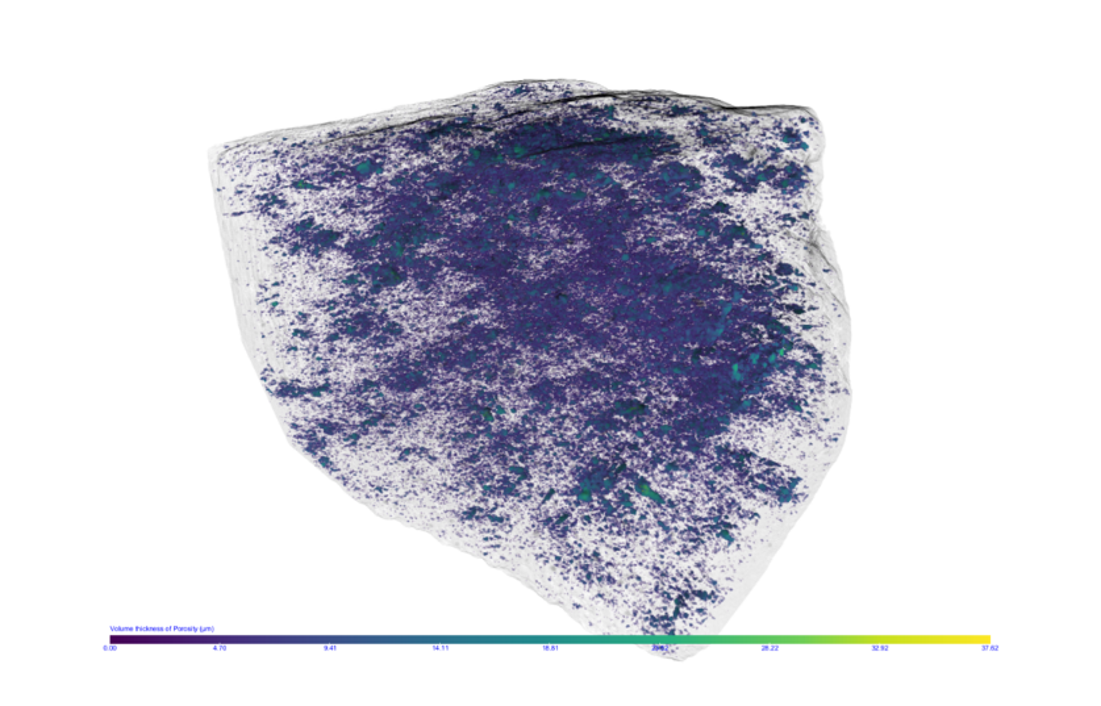
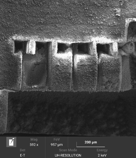
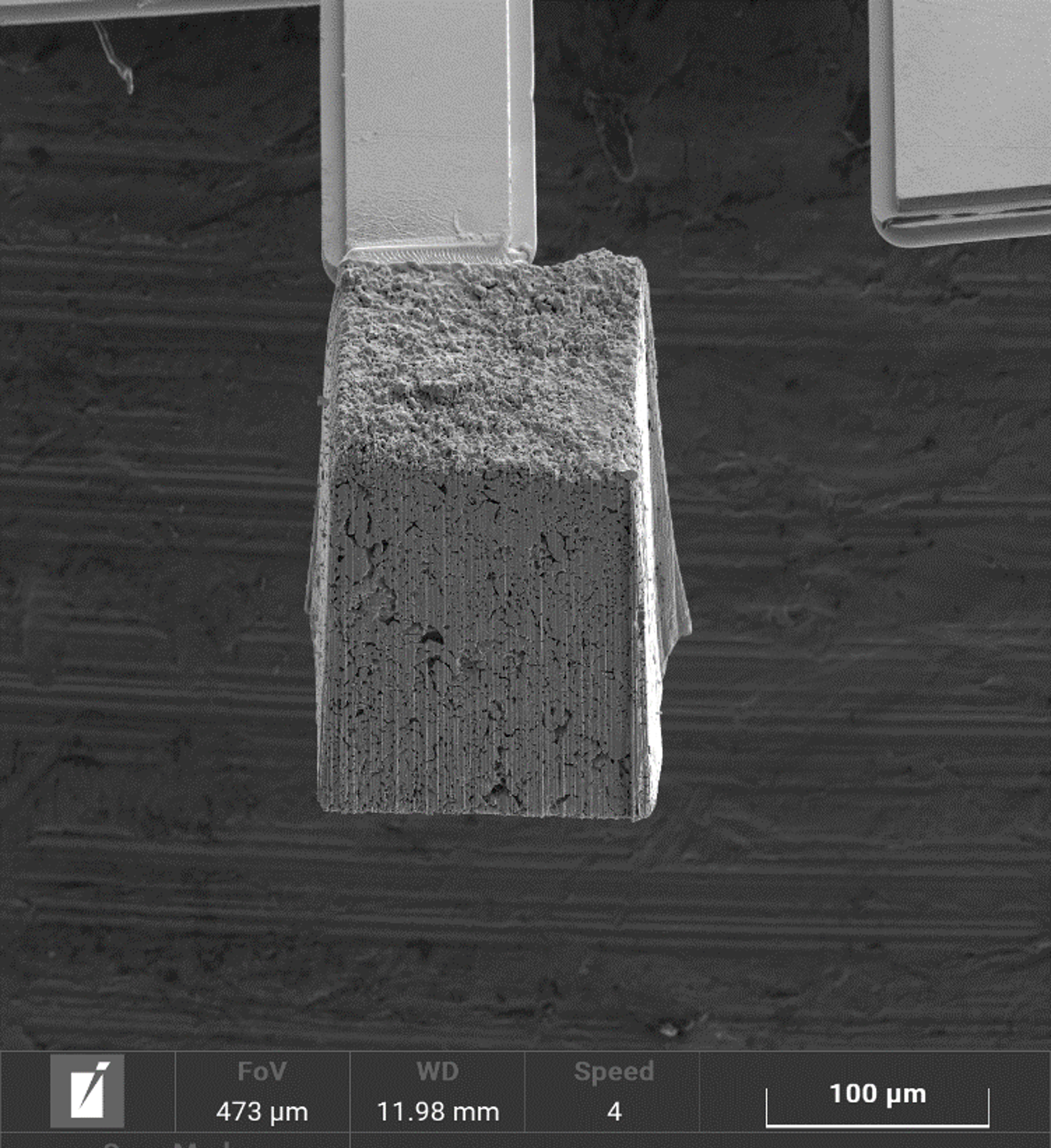
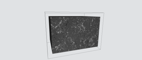
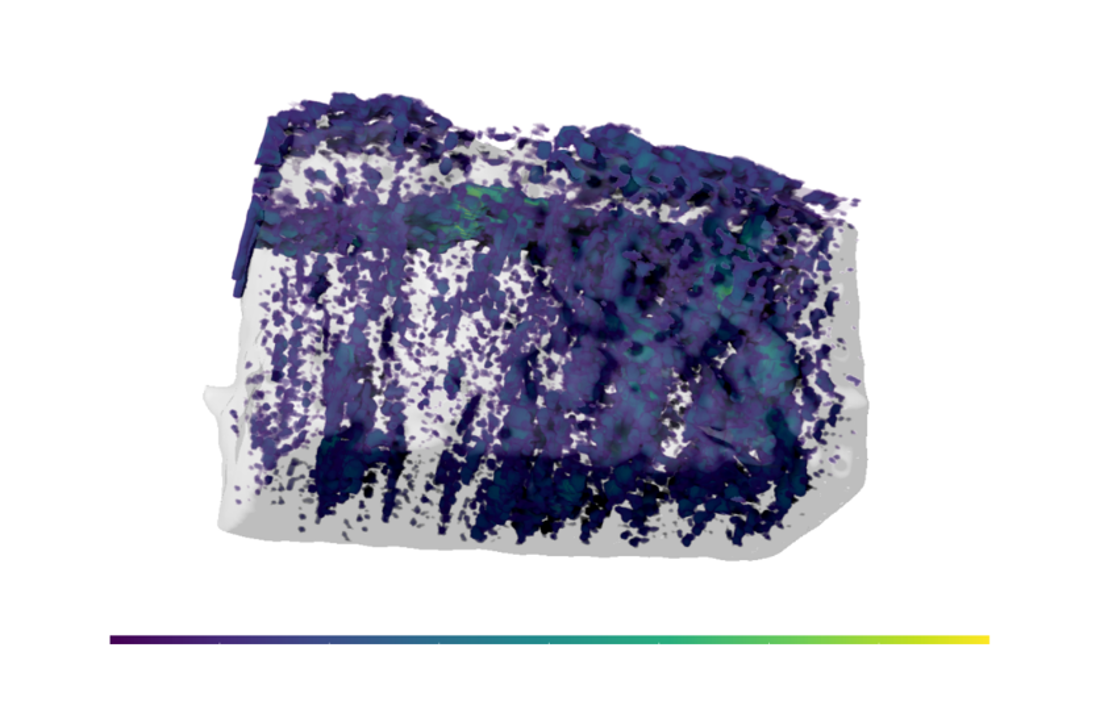
  • Enables large-volume sample prep with high-resolution FIB-SEM imaging to reveal interface detail

  • Field-free SEM provides excellent contrast at ceramic fiber boundaries, even in beam-sensitive materials

  • Seamlessly integrates into multiscale 3D tomography workflows for advanced materials analysis

AMBER-X 2

GET IN Touch

Contact us

map

Where can you find us:

Tescan
Libušina třída 21
623 00 Brno
Czech Republic

info@Tescan.com