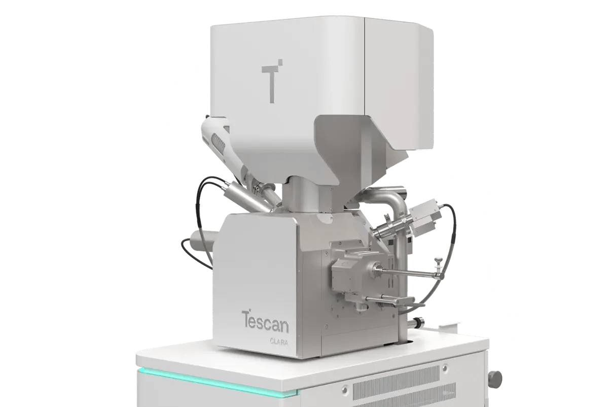
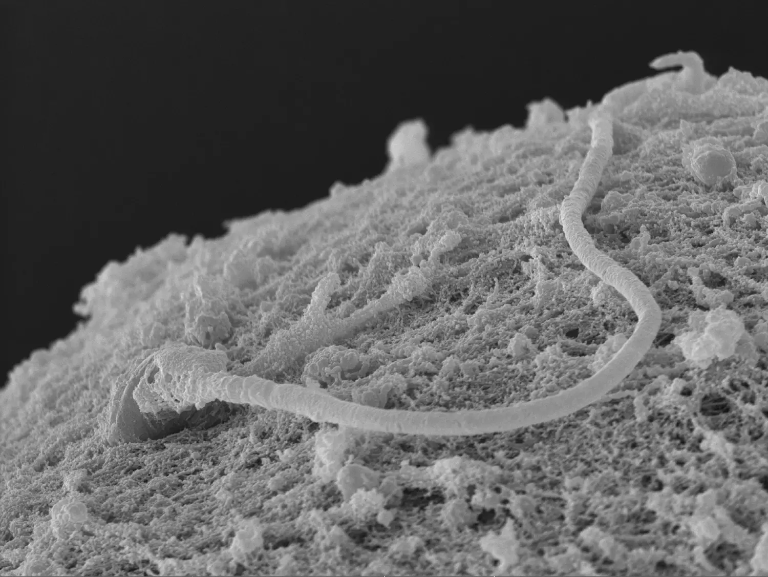
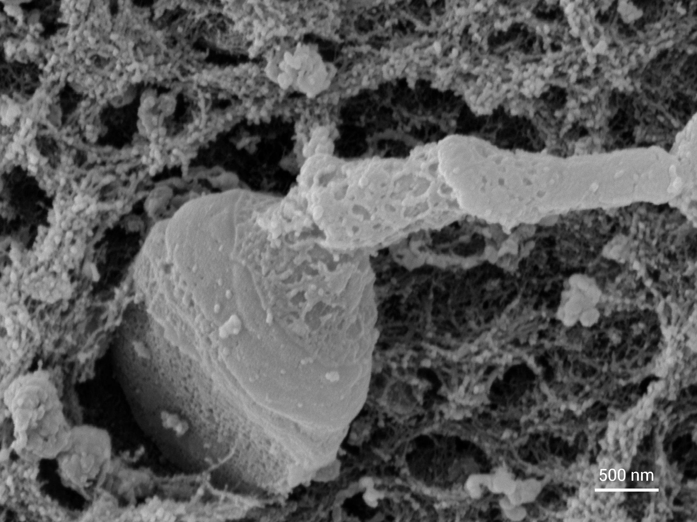
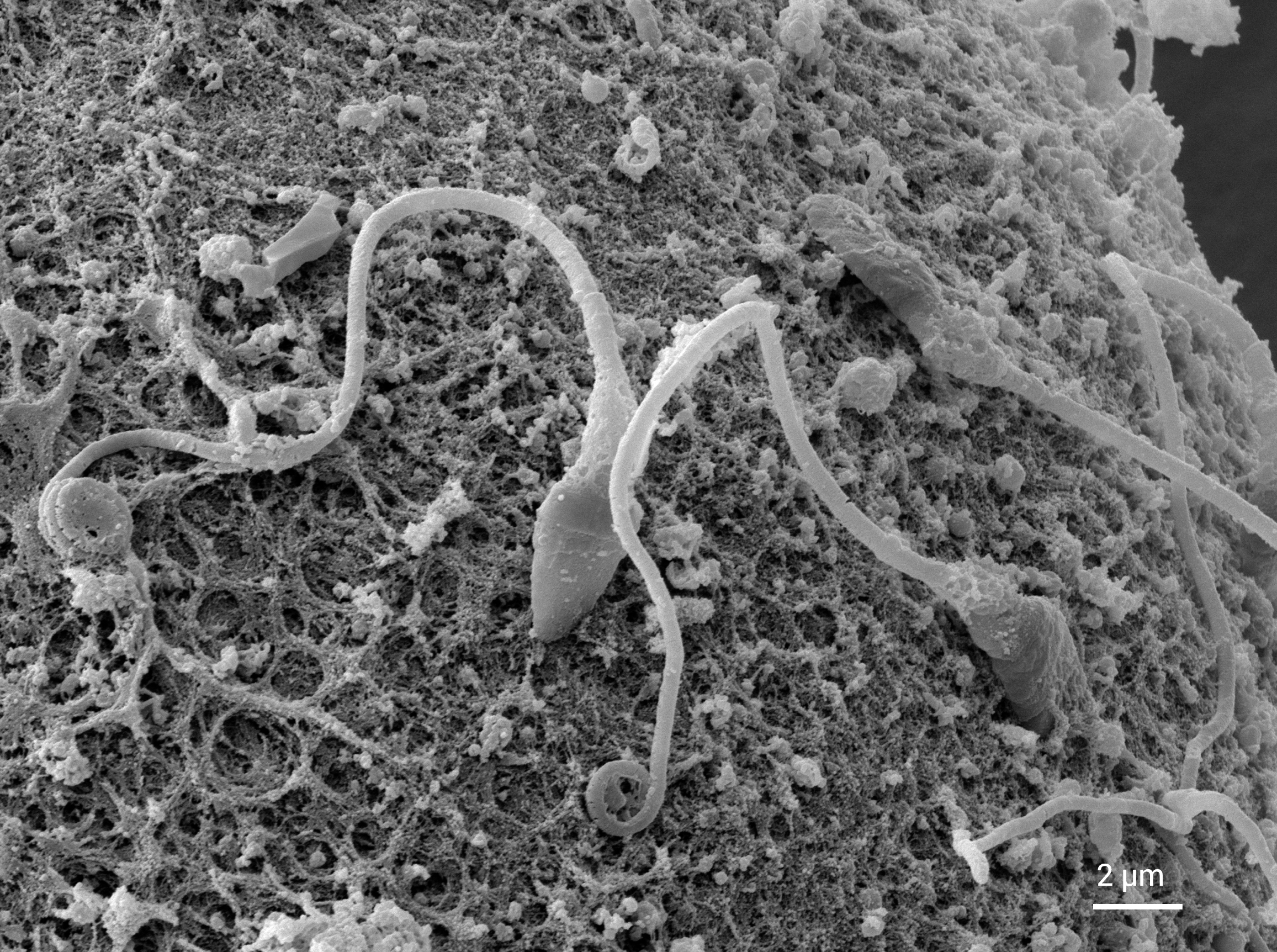
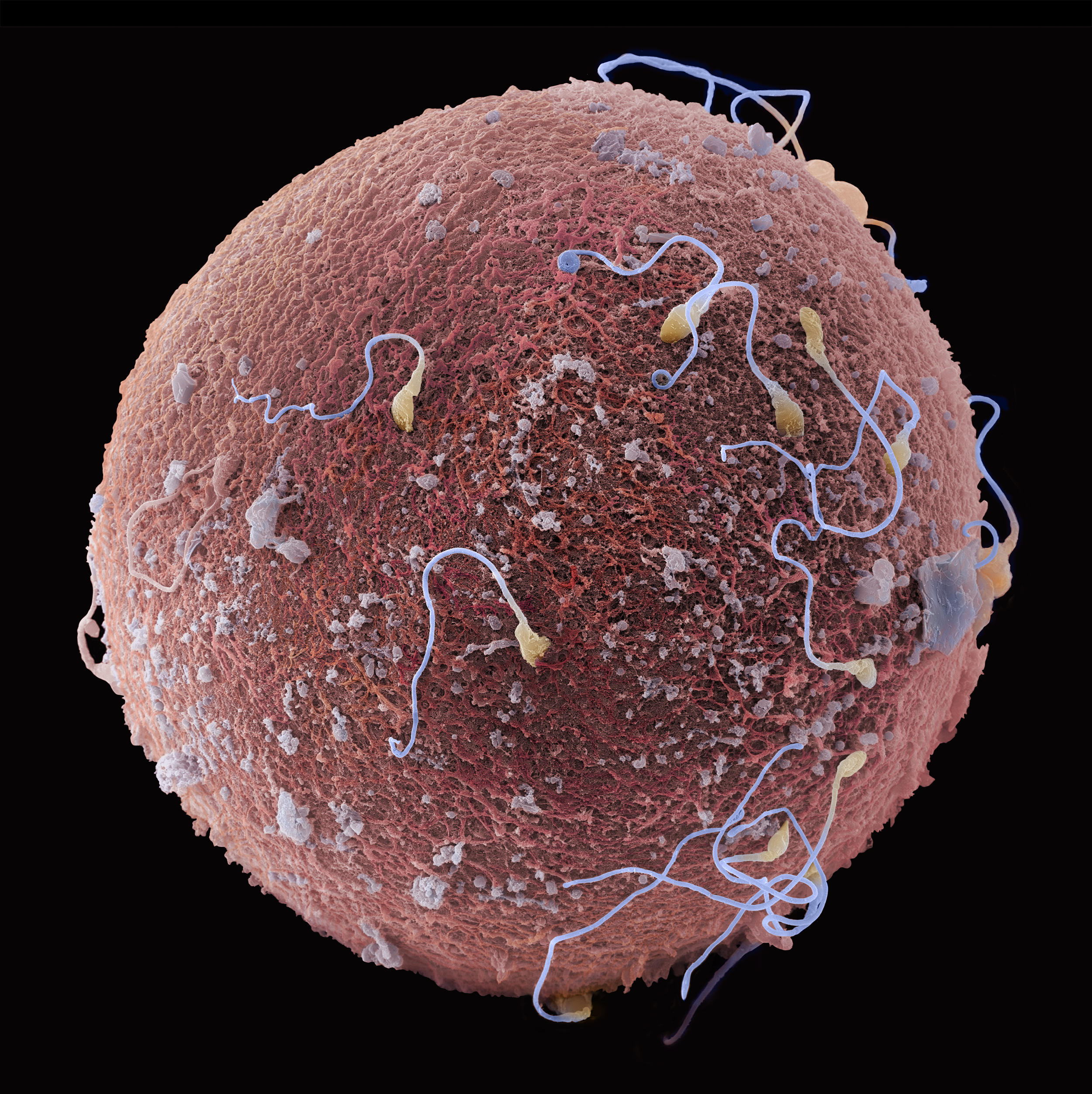
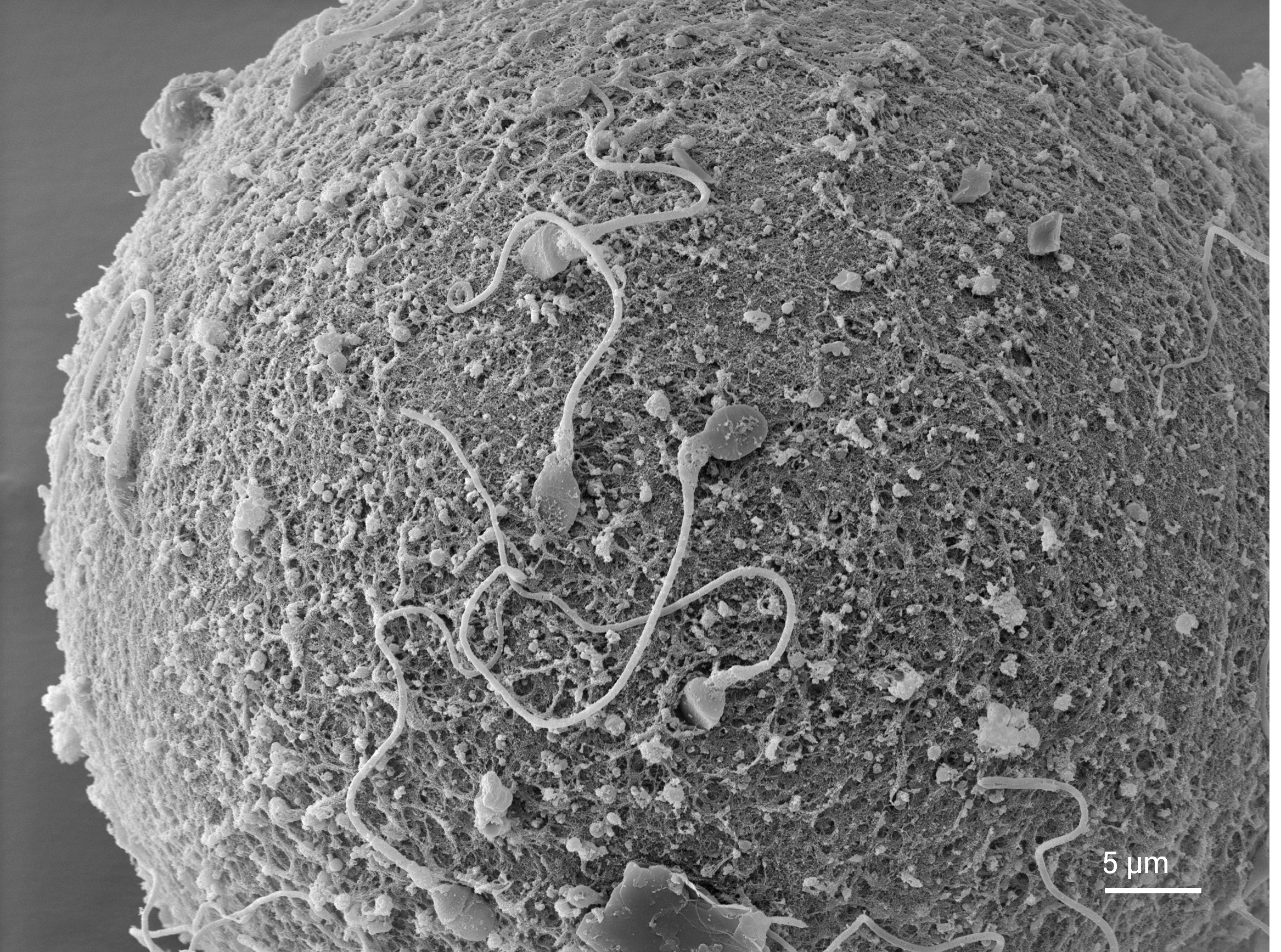
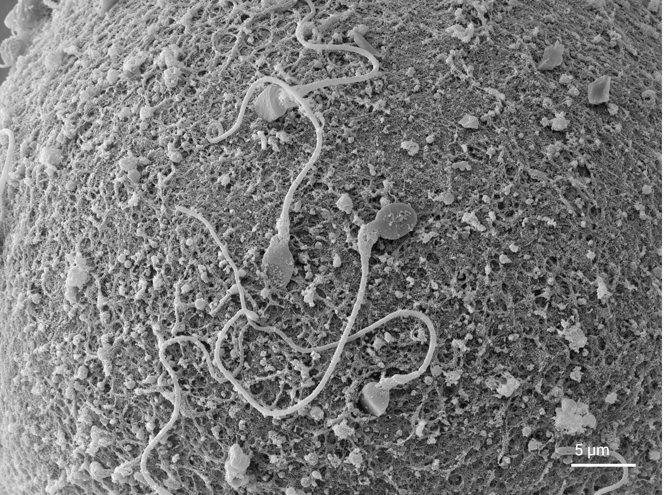
WEBINAR | From Infrastructure to Impact: EM & Micro-CT in Materials Core Facilities

Gain Insight into Human Oocyte Surface Morphology with Tescan CLARA SEM

Analyze human oocyte surface morphology and sperm interaction in ultrastructural detail using SEM imaging.

3-3-Human oocyte
3-3-Human oocyte-mobile
Unlocked content

Advancing Reproductive Research Through High-Resolution SEM Imaging

Understanding the ultrastructure of human oocytes is critical for advancing assisted reproductive technologies, yet this remains a challenge due to their fragile nature and complex surface topography. A critical step in fertilization is the interaction of sperm with the zona pellucida (the glycoprotein layer that surrounds the oocyte). This process involves intricate sperm–oocyte surface interactions that are difficult to capture in detail using conventional light microscopy.

Advanced imaging methods are therefore essential. Tescan scanning electron microscopes (SEMs) provide ultra-high resolution imaging of surface morphology and interactions such as sperm attachment, delivering insights that go beyond the limits of light microscopy. These capabilities support both fundamental research and the development of clinical applications in reproductive medicine.

Why Study Human Oocyte Surface Morphology

with TESCAN CLARA SEM?

01
Root of the Problem

Studying the surface morphology of human oocytes is essential for understanding the process of fertilization. However, light microscopy imaging techniques lack the resolution and contrast required for detailed oocyte surface analysis.

Due to complex topography, dedicated DEPTH mode was used to capture fine structural details of the egg-sperm interaction to completely visualize all details with high depth of focus.

Using both in-chamber and in-beam secondary electron (SE) detectors provided a complementary imaging advantage that is especially important for delicate, complex samples like human oocytes.

Visualize oocyte surface morphology and gamete interaction with clarity and structural integrity using Tescan SEM Systems.

02
Materials and Methods

Visualizing Human Oocyte Surface Morphology

Spare human gametes, donated from IVF treatments, were chemically fixed and stained, dehydrated, coated with gold, and prepared for high-resolution SEM imaging.

High-resolution imaging was performed using the Tescan CLARA UHR-SEM under high vacuum conditions. Due to the topographically complex surface of oocytes, a dedicated DEPTH mode was employed to maintain sharp focus across the entire surface.

Images were captured using the In-chamber secondary electron (SE) detector, the In-beam SE detector, or a combination of both  . The in-chamber detector emphasizes overall surface topography, while the in-beam detector provides high-resolution detail at short working distances. By combining signals from both detectors, we obtained comprehensive images with high resolution and accurate contrast.

This approach enabled detailed visualization of sperm–oocyte interactions, including multiple sperm heads and acrosomal remnants, vesicle structures containing enzymes critical for oocyte penetration.

03
Results and Discussion

High-Resolution Imaging of Oocyte Surface Morphology and Sperm Interaction

Ultra-high resolution SEM revealed the complex surface of human oocytes and detailed sperm-oocyte interactions, including acrosomal remnants critical for fertilization.

Using both in-chamber and in-beam SE detectors provided simultaneous topographic context and fine structural detail, preserving delicate structures while delivering ultra-high resolution, high-contrast images.

This approach reveals ultrastructural details far beyond the reach of light microscopy, enabling detailed analysis of gamete interactions, and unlocking new potential for both basic research and reproductive medicine.

GET IN Touch

Contact us

Get the most out of Tescan

This is more than information; it's an advantage. We've compiled our technical whitepapers, detailed product flyers, and on-demand webinars to provide you with the knowledge that makes a real difference. Sign up now to access the insights you need to make an impact.

Tescan Instruments & Technology

Used in This Workflow

Tescan CLARA

Tescan CLARA is engineered for advanced biological imaging under both ambient and cryogenic conditions. It supports a wide range of workflows, enabling researchers to capture complex biological structures and visualize them in 2D and 3D with exceptional detail. 

  • BrightBeam™ technology delivers ultra-high resolution and sharp, contrasted images even at very low beam energies. 

  • In-Flight™ beam tracing and automated alignments enable users to optimize imaging and analysis quickly and consistently. 

  • Wide Field Optics™ allows distortion-free navigation from 1× overview down to nanoscale features 

  • MultiVac™ Imaging Mode automates low-vacuum aperture and water-vapor operation for imaging of sensitive samples. 

  • TESCAN Essence™ interface ensures effortless operation and multi-user support for streamlined operation across all experience levels.

CLARA GM Mat. Science

Book a demo

Built around your needs

map

Where can you find us:

Tescan
Libušina třída 21
623 00 Brno
Czech Republic

130405923 us US 37.09024 -95.712891 25.3575 29.349345 20.67957527 42.082797 39.91384763 -33.693421 13.93320106 3.039986586 31.997988 38.050985 47.579533 48.1485965 58.375799 54.663142 19.195447 56.975106 50.493053 45.868592 10.79556993 44.35660598 43.2371604 55.536415 14.557577179752773 32.100937 -6.116829 -6.212299277967318 23.7104 -33.471062 31.998740087 -23.69149395 43.462349 51.529848 49.1893523 49.197486 25.072375 31.075811 1.299027 40.676979 52.30150662 51.013813 35.684121 37.479653 52.246622 40.581349 39.911632 -26.1811371 41.818215 33.429928 -12.08688

No distributors found.