

Tescan UniTOM® XL
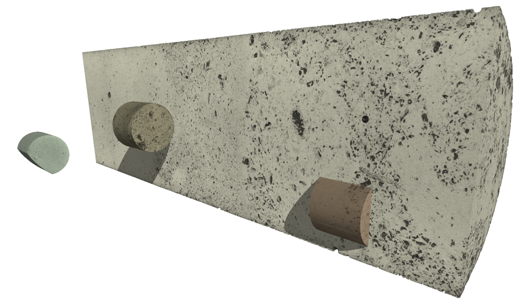
Large-volume dynamic imaging for reservoir core samples.
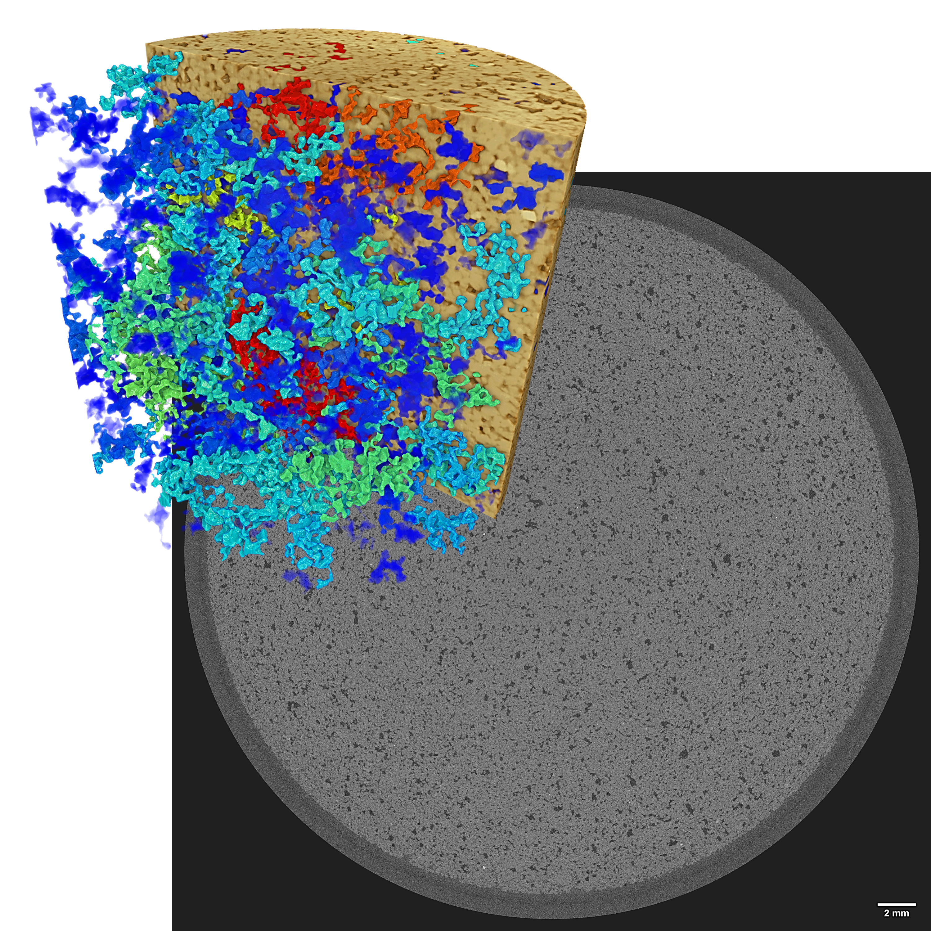
Tescan UniTOM® XL provides high-throughput, non-destructive imaging of full core plugs and large fragments.
-
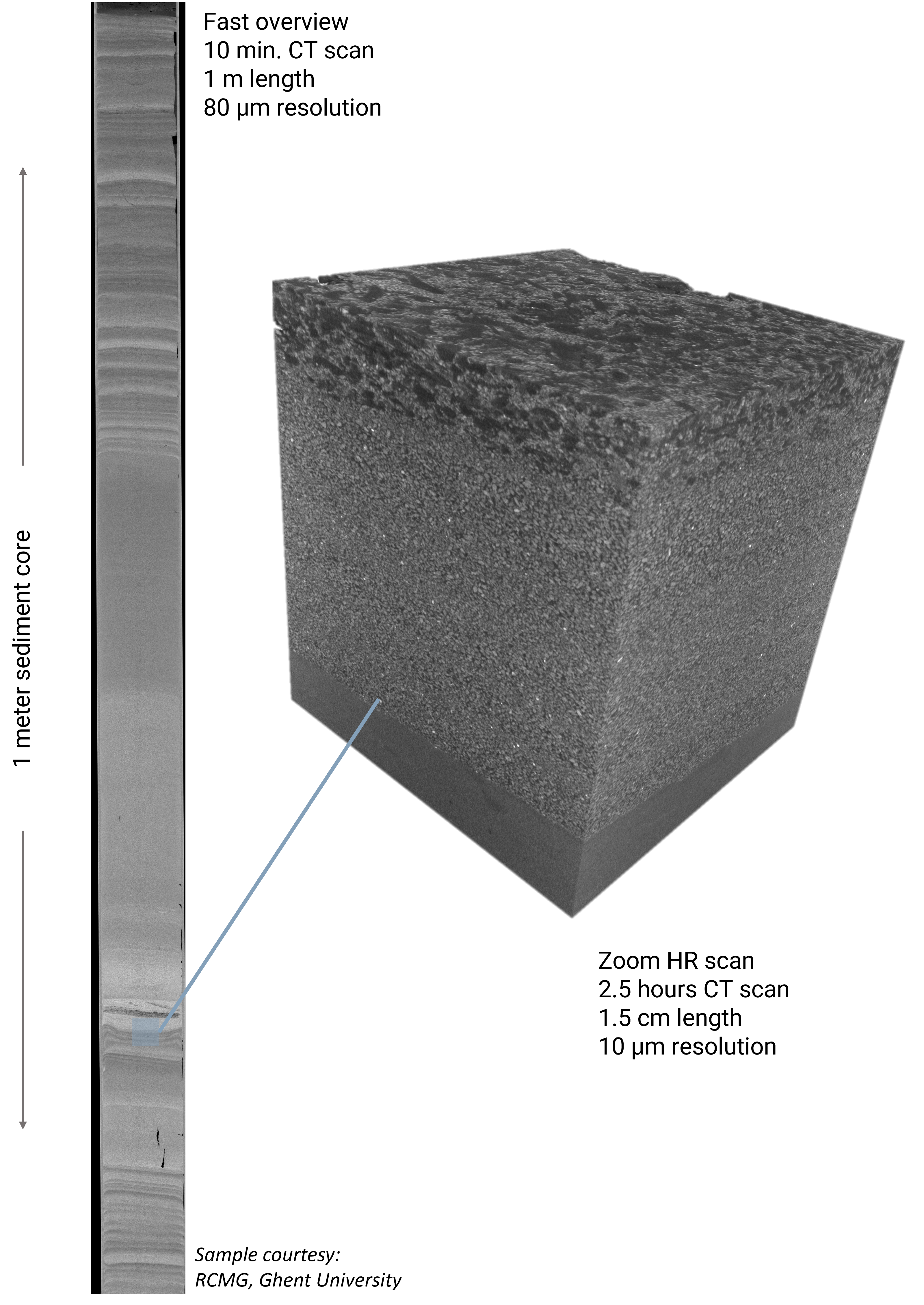
High-speed scanning with customizable field of view
-
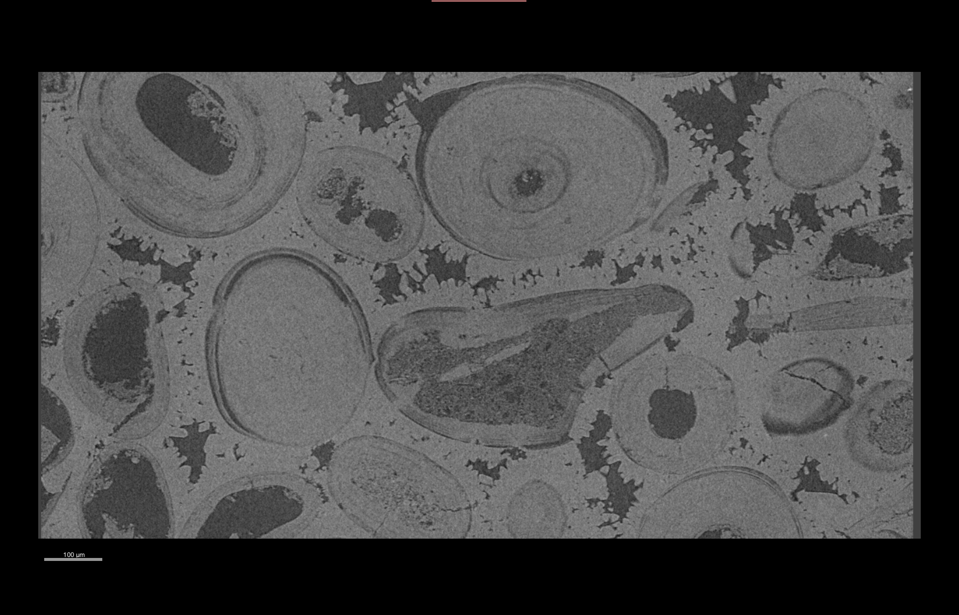
Ideal for visualizing pores and fractures across entire samples
-
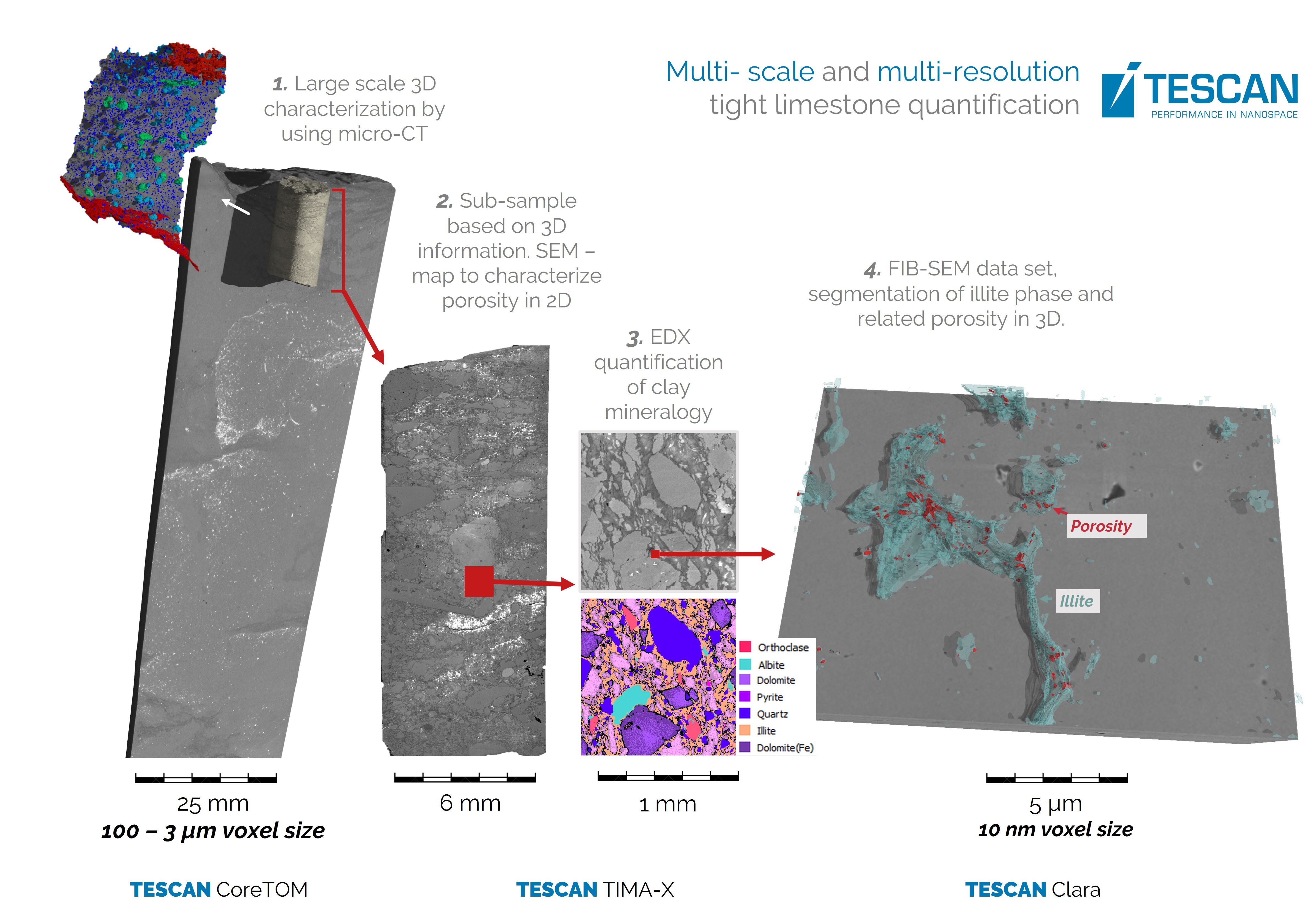
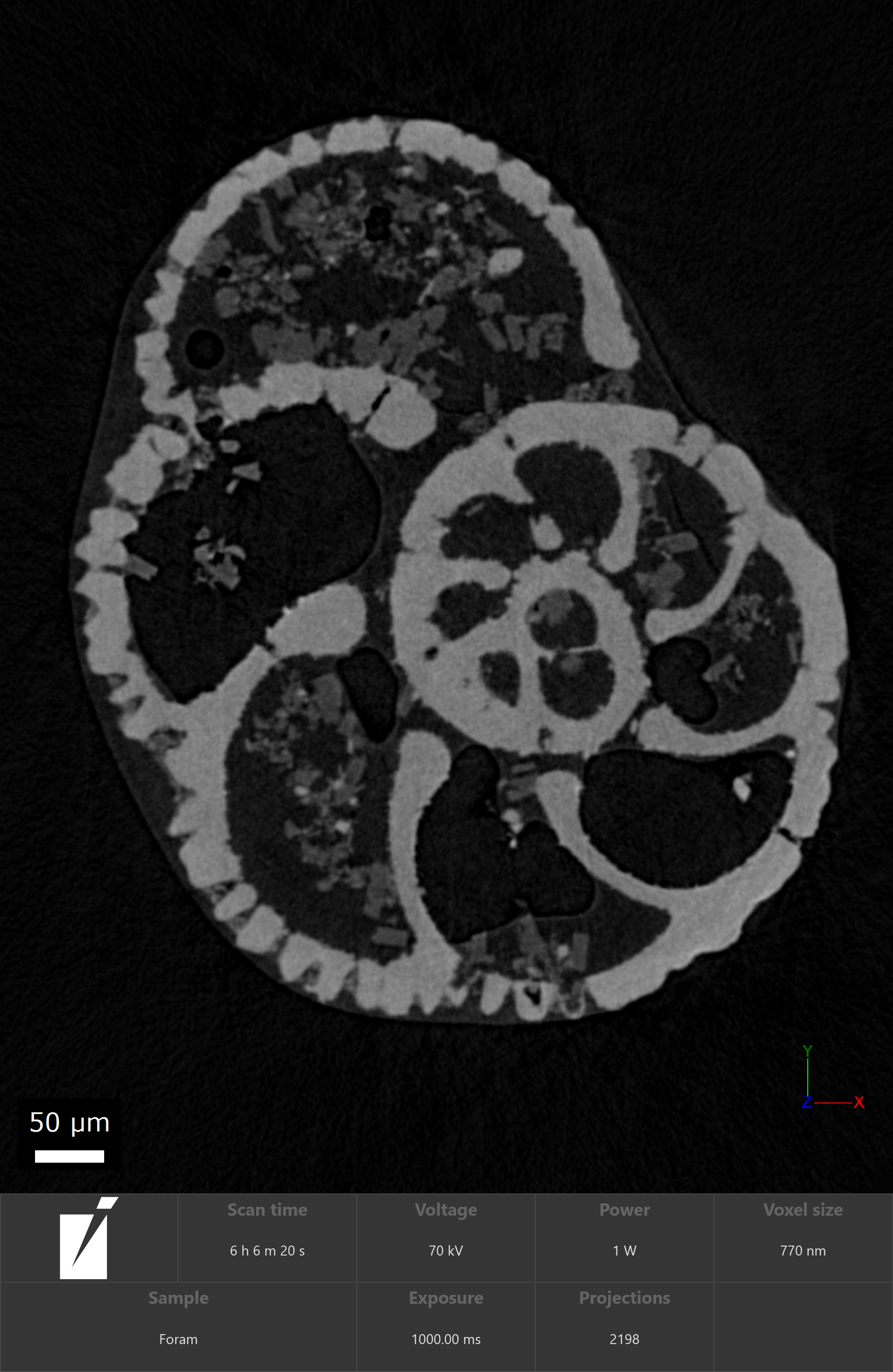
Supports integrated multiscale workflows


-1.png?width=1920&height=1211&name=2_Dynamic%20CT%20of%20multiphase%20flow.%20Pore%20filling%20events%20and%20event%20size%20are%20colorcoded%20as%20a%20function%20of%20time%20(blue=beginning%2c%20red=end)-1.png)
