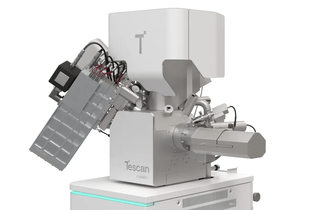
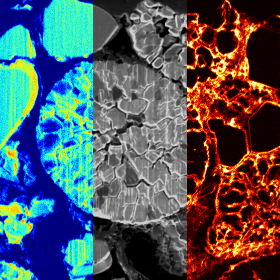
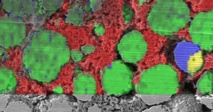
WEBINAR | Up to 40% Faster Automated TEM Lamella Preparation with new Ga⁺ FIB Column

Visualizing Electrode Particle Degradation in Lithium-Ion Batteries with Tescan AMBER X 2

Multimodal characterization using FIB-SEM integrated with ToF-SIMS and Raman spectroscopy reveals the microstructural and chemical mechanisms behind capacity loss and internal resistance growth.

hero
hero
Unlocked content

How Degraded Electrode Particles Undermine Battery Performance

Driven by cycling, heat, and side reactions, electrode particle degradation is one of the leading causes of capacity loss and internal resistance growth in lithium-ion batteries. Graphite anodes exfoliate, cathode particles fracture, and lithium inventory is lost.

In this application note, Tescan demonstrates how AMBER X 2 Plasma FIB-SEM, equipped with STEM, ToF-SIMS, and Raman spectroscopy, enables precise characterization of structural and chemical changes in both anode and cathode materials. These insights support strategies for longer battery life and safer energy storage.

Why Study Electrode Degradation

with Tescan?

01
Root of the Problem

Why Electrode Degradation Limits Battery Lifespan

Every charge and discharge cycle subtly alters the microstructure of electrode particles. Graphite anodes exfoliate as lithium intercalates between layers. Over time, this creates internal gaps and increases resistance. On the cathode side, NMC particles crack under mechanical and chemical stress, fragmenting and losing contact.

These failures not only reduce energy capacity but also promote side reactions and loss of lithium inventory. Because these processes occur at the micro- and nanoscale, precise tools are needed to study them in detail — both structurally and chemically.

02
Materials and Methods

Multimodal FIB-SEM and Correlative Chemical Analysis

Electrode degradation was studied in commercial graphite anodes and NMC cathodes using the Tescan AMBER X 2 Plasma FIB-SEM system. Samples were transferred from the glovebox via inert gas transfer. Imaging techniques included SEM, STEM, 3D FIB-SEM tomography, and 3D ToF-SIMS tomography.

Raman spectroscopy was also applied, all within the same platform. Lamellae for cross-sectional and TEM-level analysis were prepared in situ. This multimodal workflow enabled full correlation between surface features, internal microstructures, and chemical composition — information critical for degradation analysis.

03
Results and Discussion

Exfoliation, Fracturing, and Lithium Trapping Visualized in 3D

Graphite anodes showed clear exfoliation on both the surface and in TEM lamellae. 3D FIB-SEM and ToF-SIMS tomography revealed that lithium-based compounds accumulate in these regions, confirming loss of lithium inventory and active materials due to graphite particle degradation. NMC cathodes, analyzed via cross section and Raman mapping, showed fractured particles and clear signs of inhomogeneous cycling behavior.

Raman spectra revealed peak broadening and shifts indicating internal stress and non-uniform lithium distribution. Some particles were inactive — not participating in cycling — further reducing capacity. These results confirm the value of multimodal analysis for guiding material improvements and battery lifespan extension strategies.

GET IN Touch

Contact us

Get the most out of Tescan

This is more than information; it's an advantage. We've compiled our technical whitepapers, detailed product flyers, and on-demand webinars to provide you with the knowledge that makes a real difference. Sign up now to access the insights you need to make an impact.

Tescan Instruments & Technology

Used in This Workflow

Tescan AMBER X 2

Tescan AMBER X 2 combines plasma FIB milling with ultra-high-resolution SEM and integrated chemical analysis tools — ideal for studying electrode materials at every scale.

  • Plasma FIB for fast lamella milling and volume sectioning

  • Field-free UHR SEM with low-kV imaging for surface-sensitive features

  • Integrated RSTEM for nanometer-scale subsurface analysis

  • Compatible with inert sample transfer workflows 
AMBER-X2

Tescan ToF-SIMS and Raman Spectroscopy – Integrated Chemical Mapping Tools

Integrated into the AMBER X 2 platform, these tools reveal material composition, lithium distribution, and cycling-induced changes.

  • 3D ToF-SIMS tomography identifies lithium compounds inside degraded regions

  • Raman spectroscopy maps structural disorder and inactive particles

  • Enables correlation of morphology, defect sites, and chemical evolution 

GET IN Touch

Contact us

map

Where can you find us:

Tescan Brno
Libušina třída 21
623 00 Brno
Czech Republic

info@Tescan.com